

Advances in retinal stem cell biology
- PMID: 23943690
- PMCID: PMC3740467
Advances in retinal stem cell biology
Abstract
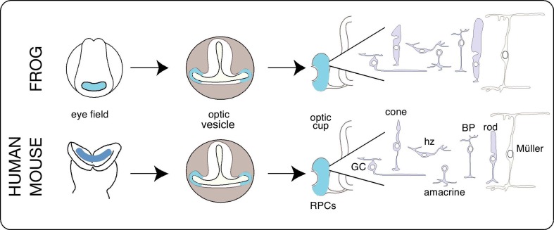
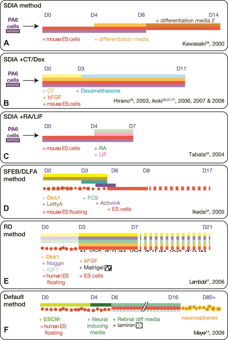
Tremendous progress has been made in recent years to generate retinal cells from pluripotent cell sources. These advances provide hope for those suffering from blindness due to lost retinal cells. Understanding the intrinsic genetic network in model organisms, like fly and frog, has led to a better understanding of the extrinsic signaling pathways necessary for retinal progenitor cell formation in mouse and human cell cultures. This review focuses on the culture methods used by different groups, which has culminated in the generation of laminated retinal tissue from both embryonic and induced pluripotent cells. The review also briefly describes advances made in transplantation studies using donor retinal progenitor and cultured retinal cells.
Keywords: Artificial Retina; Cone Photoreceptors; Embryonic Stem Cells, ES; Eye; Ganglion Cells; Genetic Network; Induced Pluripotent Stem Cells, iPS; Noggin; Retina; Signaling Pathways; Stromal Cells.
Figures
References
-
- Müller F, O’Rahilly R. The first appearance of the neural tube and optic primordium in the human embryo at stage 10. Anat Embryol (Berl) 1985;172:157–169. - PubMed
-
- Zuber ME, Harris WA. Formation of the eye field. In: Sernagor E, Eglen S, Harris WA, Wong R, editors. Retinal Development. Cambridge: Cambridge University Press; 2006. pp. 8–29.
-
- Lanza RP. Essentials of stem cell biology. Boston: Elsevier/Academic Press; 2009.
-
- Amato MA, Arnault E, Perron M. Retinal stem cells in vertebrates: parallels and divergences. Int J Dev Biol. 2004;48:993–1001. - PubMed
Grants and funding
LinkOut - more resources
Full Text Sources