

A case of orbital emphysema associated with frontal sinus pneumocele
- PMID: 23943722
- PMCID: PMC3713563
- DOI: 10.1055/s-0033-1347903
A case of orbital emphysema associated with frontal sinus pneumocele
Abstract
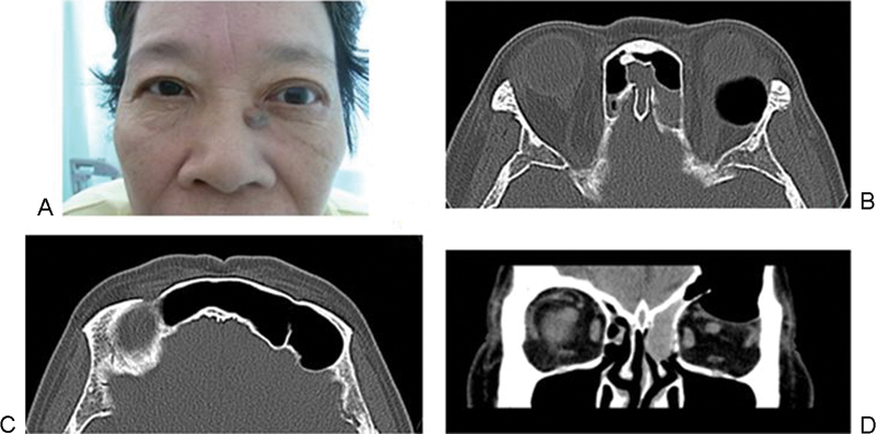
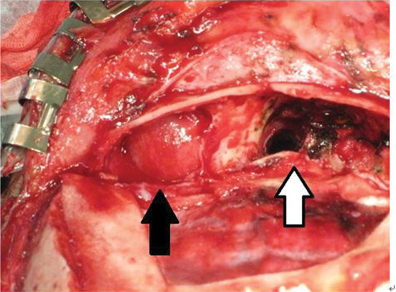
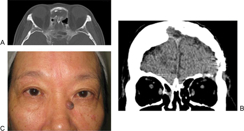
Orbital emphysema is usually caused by trauma and fracture of an orbital bone, allowing air to pass from the sinuses into the orbit. Orbital emphysema without any significant trauma is rare. We present a case of a 67-year-old-woman who complained of left exophthalmos without any history of trauma, sneezing, or sinus surgery. Computed tomography scanning showed left orbital emphysema protruding the eyeball forward. The left frontal sinus was remarkably enlarged associated with a partial defect of the orbital roof, allowing air entry into the orbit. In addition, the frontal sinus ostium was occluded with the mucocele that served as a one-way valve between the frontal and the ethmoidal sinuses. We performed frontal craniotomy and removed the mucocele and the inner table of frontal bone to communicate the frontal sinus with the nasal cavity. After operation, her exophthalmos was improved.
Keywords: frontal sinus pneumocele; orbital emphysema; transcranial surgery.
Figures
Similar articles
-
Pneumocele of the frontal sinus producing orbital roof defect: case report and review of literature.Am J Otolaryngol. 2010 May-Jun;31(3):202-4. doi: 10.1016/j.amjoto.2009.01.001. Epub 2009 Apr 23. Am J Otolaryngol. 2010. PMID: 20015740 Review.
-
Frontal mucocele with an accompanying orbital abscess mimicking a fronto-orbital mucocele: case report.BMC Ear Nose Throat Disord. 2006 Apr 18;6:6. doi: 10.1186/1472-6815-6-6. BMC Ear Nose Throat Disord. 2006. PMID: 16620377 Free PMC article.
-
Orbital complications of infected mucocele in the paranasal sinuses.Auris Nasus Larynx. 2020 Dec;47(6):990-995. doi: 10.1016/j.anl.2020.05.012. Epub 2020 Jun 11. Auris Nasus Larynx. 2020. PMID: 32536502
-
Pneumocele--a rare cause of air in the orbit.Am J Ophthalmol. 2004 Jul;138(1):168-9. doi: 10.1016/j.ajo.2004.02.054. Am J Ophthalmol. 2004. PMID: 15234310
-
Osteoplasty flap technique for repair of latent (30-year) post-traumatic frontal sinus mucocele: case report and review of the literature.J Oral Maxillofac Surg. 2012 Sep;70(9):2092-6. doi: 10.1016/j.joms.2011.10.015. Epub 2012 Apr 26. J Oral Maxillofac Surg. 2012. PMID: 22542331 Review.
Cited by
-
Frontal sinus pneumocele caused by a maxillary sinus mucocele.BMJ Case Rep. 2021 Apr 23;14(4):e242477. doi: 10.1136/bcr-2021-242477. BMJ Case Rep. 2021. PMID: 33893136 Free PMC article.
-
Rare Diseases of the Nose, the Paranasal Sinuses, and the Anterior Skull Base.Laryngorhinootologie. 2021 Apr;100(S 01):S1-S44. doi: 10.1055/a-1331-2469. Epub 2021 Apr 30. Laryngorhinootologie. 2021. PMID: 34352902 Free PMC article. Review.
References
-
- Zimmer-Galler I E, Bartley G B. Orbital emphysema: case reports and review of the literature. Mayo Clin Proc. 1994;69(2):115–121. - PubMed
-
- Mensiz E, Tüz M, Oyar O, Doğru H, Yasan H. A case of orbital emphysema associated with internal laryngocele. Auris Nasus Larynx. 2003;30(2):197–200. - PubMed
-
- Boulos P R, Bernardino C R, Rubin P AD. Pneumocele—a rare cause of air in the orbit. Am J Ophthalmol. 2004;138(1):168–169. - PubMed
LinkOut - more resources
Full Text Sources
Other Literature Sources