. 2013 Oct;6(10):1128-39.
doi: 10.1158/1940-6207.CAPR-13-0209. Epub 2013 Aug 13.

Acacetin inhibits in vitro and in vivo angiogenesis and downregulates Stat signaling and VEGF expression

Affiliations

Acacetin inhibits in vitro and in vivo angiogenesis and downregulates Stat signaling and VEGF expression

Tariq A Bhat et al. Cancer Prev Res (Phila). 2013 Oct.

Abstract

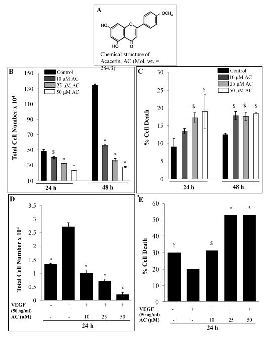
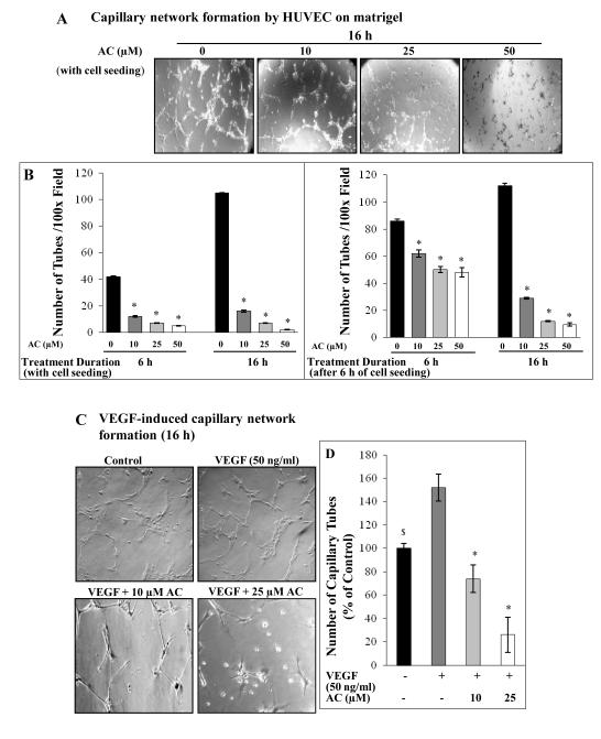
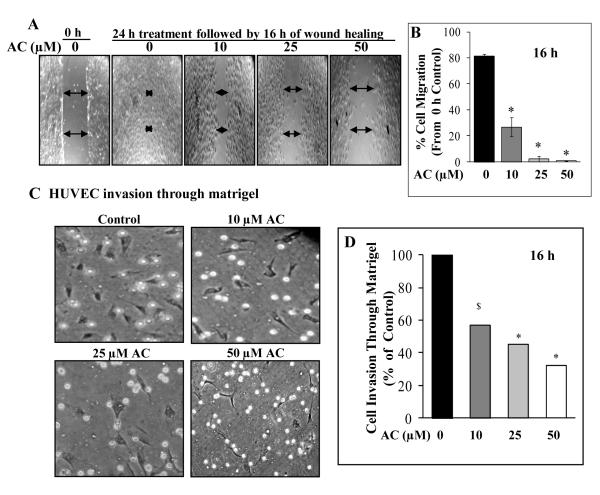
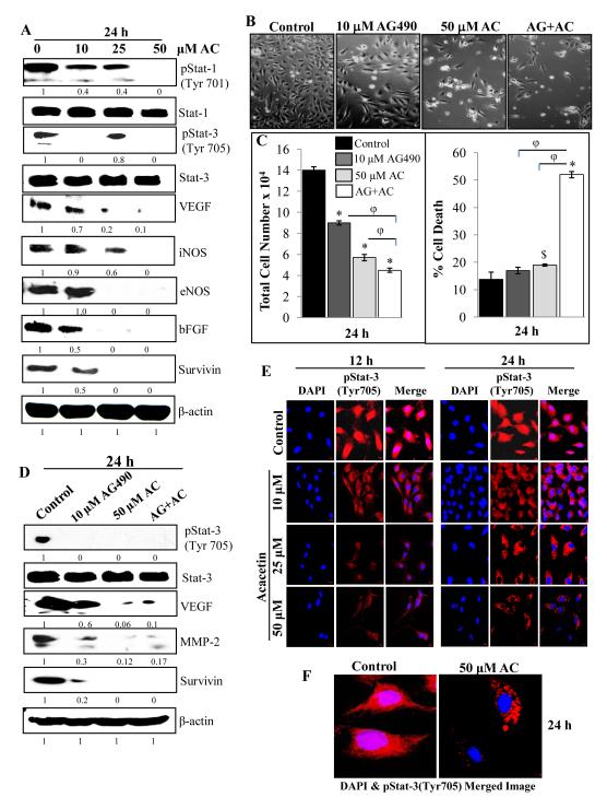
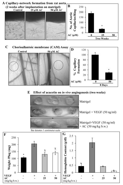
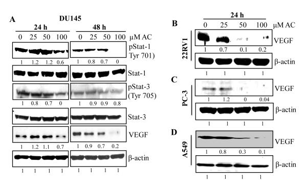
Angiogenesis is an effective target in cancer control. The antiangiogenic efficacy and associated mechanisms of acacetin, a plant flavone, are poorly known. In the present study, acacetin inhibited growth and survival (up to 92%; P < 0.001), and capillary-like tube formation on Matrigel (up to 98%; P < 0.001) by human umbilical vein endothelial cells (HUVEC) in regular condition, as well as VEGF-induced and tumor cells conditioned medium-stimulated growth conditions. It caused retraction and disintegration of preformed capillary networks (up to 91%; P < 0.001). HUVEC migration and invasion were suppressed by 68% to 100% (P < 0.001). Acacetin inhibited Stat-1 (Tyr701) and Stat-3 (Tyr705) phosphorylation, and downregulated proangiogenic factors including VEGF, endothelial nitric oxide synthase (eNOS), inducible nitric oxide synthase (iNOS), matrix metalloproteinase-2 (MMP-2), and basic fibroblast growth factor (bFGF) in HUVEC. It also suppressed nuclear localization of pStat-3 (Tyr705). Acacetin strongly inhibited capillary sprouting and networking from rat aortic rings and fertilized chicken egg chorioallantoic membrane (CAM; ∼71%; P < 0.001). Furthermore, it suppressed angiogenesis in Matrigel plugs implanted in Swiss albino mice. Acacetin also inhibited tyrosine phosphorylation of Stat-1 and -3, and expression of VEGF in cancer cells. Overall, acacetin inhibits Stat signaling and suppresses angiogenesis in vitro, ex vivo, and in vivo, and therefore, it could be a potential agent to inhibit tumor angiogenesis and growth.


Figures

Figure 1
Figure 1
Effect of acacetin on HUVEC growth and proliferation. A: Chemical structure of acacetin (AC). B-C: HUVEC proliferation and death after 24 and 48 h of AC treatment. Cells were grown in complete EGM-2MV media with 5% FBS at the density of 1× 10⁵ cells/60 mm culture plates. After 24 h of seeding, cells were treated with 10-50 μM concentrations of AC for 24-48 h in regular growth conditions. At the end of the experiment, cells were harvested and counted using trypan blue as mentioned in Methods. Total cell number and percent dead cells versus control are shown. D-E: Effect of acacetin on VEGF-stimulated HUVEC proliferation and death. HUVEC were cultured at the density of 20,000 cells/12-well plates for 24 h time followed by serum-starvation overnight before treatment with the indicated doses of AC in serum-free medium supplemented with or without VEGF for 24 h as discussed in Methods. Total cell number and percent dead cells versus control are shown. The quantitative data shown are Mean ± SE of three samples for each treatment. Equal volumes of DMSO (0.1%, v/v) were present in each treatment. $, p<0.05; *, p<0.001 versus DMSO control or VEGF-alone treatment.
Figure 2
Figure 2
Effect of acacetin on capillary tube formation by HUVEC on matrigel. A: Representative images depicting formation of capillary tubes on matrigel by HUVEC after 16 h treatment with AC at the time of cell seeding. B: Quantitative depiction of capillary tube formation after 6 and 16 h of AC treatment either at the time of cell seeding or after 6 h of initial cell seeding. HUVEC (4× 10⁴ cells/well) were either simultaneously seeded and treated with AC or treatment was done after 6 h of initial cell seeding in 24-well matrigel-coated culture plates, and tube formation was observed and quantified periodically over a specific time period as mentioned in Methods. C: Representative images depicting formation of capillary-like tubes on matrigel by HUVEC after 16 h treatment with AC in VEGF-stimulated conditions. D: Quantitative data of the effect of AC treatment on VEGF-stimulated capillary tube formation by HUVEC. Cells were overnight starved and treated with AC and/or VEGF for 16 h as mentioned in Methods. Tubular network was photographed at 100x magnification and scored by counting the number of closed structure made by three or more independent cells under an inverted microscope. Quantitative data shown are Mean ± SE of three samples. $, p<0.05; *, p<0.001 versus DMSO or VEGF control.
Figure 3
Figure 3
Effect of acacetin on migration and invasion potential of HUVEC. A: Representative images depicting cell migration by HUVEC after 16 h following 24 h treatment with or without AC in the wound healing assay. Wound closure was recorded at 0 and 16 h after injury using an inverted microscope equipped with a digital camera as mentioned in Methods. B: Bar diagram showing the effect of 24 h AC treatment on wound closure/migration potential of HUVEC after 16 h post injury. Five independent areas in each wound were measured. Data are shown as percent cell migration compared to 0 h control at the time of injury. C: Representative images and its graphical representation depicting the effect of AC (0, 10, 25 and 50 μM) on HUVEC invasion/migration in matrigel-coated Boyden chambers after 16 h treatment. Cells were allowed to invade and migrate for 16 h and the invaded/migrated cells at the bottom of the membrane were fixed, stained and counted at 200x magnification, and photographed. D: Five independent areas were scored in each sample and data are shown as percent cell migration compared to control. $, p<0.005; *, p<0.001 versus DMSO control.
Figure 4
Figure 4
Acacetin inhibits Stat signaling and expression of angiogenic factors in HUVEC. A: HUVEC were grown to 70% confluency and treated with the indicated concentrations of AC for 24 h. Whole cell lysates were analyzed by immunoblotting using specific primary antibodies for pStat-1 (Tyr701); Stat-1; pStat-3 (Tyr705); Stat-3; VEGF, iNOS, eNOS, bFGF and survivin followed by detection with HRP-labeled appropriate secondary antibodies as mentioned in Methods. β-actin was probed after stripping the membrane as protein loading control. B-C: Representative pictures and quantitative data depicting the effect of AG490 (Janus kinase inhibitor) and AC in individual as well as combination treatments on HUVEC growth and death after 24 h of treatment in regular conditions. D: In similar treatments as in (C), whole cell lysates were analyzed by immunoblotting using specific primary antibodies for pStat-3 (Tyr705); Stat-3; VEGF, MMP-2 and survivin. Membranes were stripped and re-probed for beta-actin as loading control. E: Confocal depictions of the effects of AC on nuclear translocation of pStat-3. HUVEC were grown on coverslips for 24 h followed by treatments with the indicated doses of AC for indicated time points in regular growth conditions. At the end of the treatments, cells were processed for confocal assay as mentioned in Methods using specific primary pStat-3 (Tyr705) antibody followed by incubation with Alexa Fluor 488-secondary antibody. Slides were mounted and immediately viewed and photographed under LSM780 Laser Confocal Microscope F: A higher magnification image is shown for control and 50 μM AC for 24 h treatment. $, p<0.005; *, p<0.001 versus DMSO control; φ, p<0.001 versus AC or AG490 treatment.
Figure 5
Figure 5
Effects of acacetin on ex vivo and in vivo angiogenesis. A-B: Rat aortic ring sections were embedded in matrigel and cultured in complete EGM2-MV medium. After two days, AC (0, 25 and 50 μM) treatments were started and continued after every 48 h for 2 weeks. At the end, rings were photographed and vessels were scored by counting total number of vessels originating from the rings. A: Representative pictures depicting the effect of AC on rat aortic ring angiogenesis; and B: quantitative representation of the data as Mean ± SE of total number of aortic capillaries as a function of AC concentration. C-D: For CAM angiogenesis assay, fertilized chicken eggs were incubated and treated with AC every 48 h for 8 days as mentioned in Methods. Thereafter, individual CAMs were analyzed and photographed using digital camera with 10x zoom (C) and data were quantified by counting the nascent vessels between the major blood vessels and represented as Mean ± SE of total number of vessels in 5 independent areas on CAMs for each treatment (D). E-G: Acacetin suppresses VEGF-induced in vivo angiogenesis. Mice were randomly divided into four groups (n=5/group); and subcutaneously received matrigel only (Control), or matrigel + VEGF (50 ng/ml) or matrigel + VEGF (50 ng/ml) + AC (25 or 50 mg/kg body wt). After 14 days, mice were sacrificed and plugs were retrieved, photographed, weighed and hemoglobin content measured as described in Methods. E: Photographs of representative matrigel plugs, F: Weight of matrigel plugs (mg/plug); and G: hemoglobin content (g/dl) are shown. Body weight and diet/water consumption did not change among groups (data not shown). $, p<0.005; *, p<0.001 versus DMSO control or VEGF control.
Figure 6
Figure 6
Effects of acacetin on activation and/or expression of various proangiogenic factors in human cancer cells. A-C: Human prostate carcinoma DU145, 22Rv1 and PC-3 and D: human lung carcinoma A549 cells were grown to 70% confluency and treated with the indicated concentrations of AC for 24 and/or 48 h in regular growth conditions. Subsequently, whole cell lysates were prepared and analyzed by immunoblotting using specific primary antibodies for pStat-1 (Tyr701); Stat-1; pStat-3 (Tyr705); Stat-3 and VEGF proteins followed by detection with HRP-labeled secondary antibodies using enhanced chemiluminescence detection system. β-actin was probed after stripping the membranes as protein loading control.

References

    1. Ferrara N, Kerbel RS. Angiogenesis as a therapeutic target. Nature. 2005;438:967–74. - PubMed
    1. Naumov GN, Akslen LA, Folkman J. Role of angiogenesis in human tumor dormancy: animal models of the angiogenic switch. Cell Cycle. 2006;5(16):1779–87. - PubMed
    1. Naumov GN, Folkman J, Straume O. Tumor dormancy due to failure of angiogenesis: role of the microenvironment. Clin Exp Metastasis. 2009;26(1):51–60. - PubMed
    1. Halvorsen O, Haukaas S, Hoisaeter P, Akslen L. Independent prognostic importance of microvesel density in clinically localized prostate cancer. Anticancer Res. 2000;20:3791–99. - PubMed
    1. Gettman MT, Pacelli A, Slezak J, Bergstralh EJ, Blute M, Zincke H, Bostwick DG. Role of microvessel density in predicting recurrence in pathologic Stage T3 prostatic adenocarcinoma. Urology. 1999;54:479–85. - PubMed

Publication types

MeSH terms