Review
. 2014 Nov-Dec;35(11):2030-8.
doi: 10.3174/ajnr.A3695. Epub 2013 Aug 14.

PET approaches for diagnosis of dementia

Affiliations
Review

PET approaches for diagnosis of dementia

K Ishii. AJNR Am J Neuroradiol. 2014 Nov-Dec.

Abstract

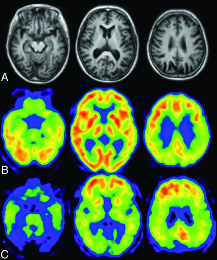
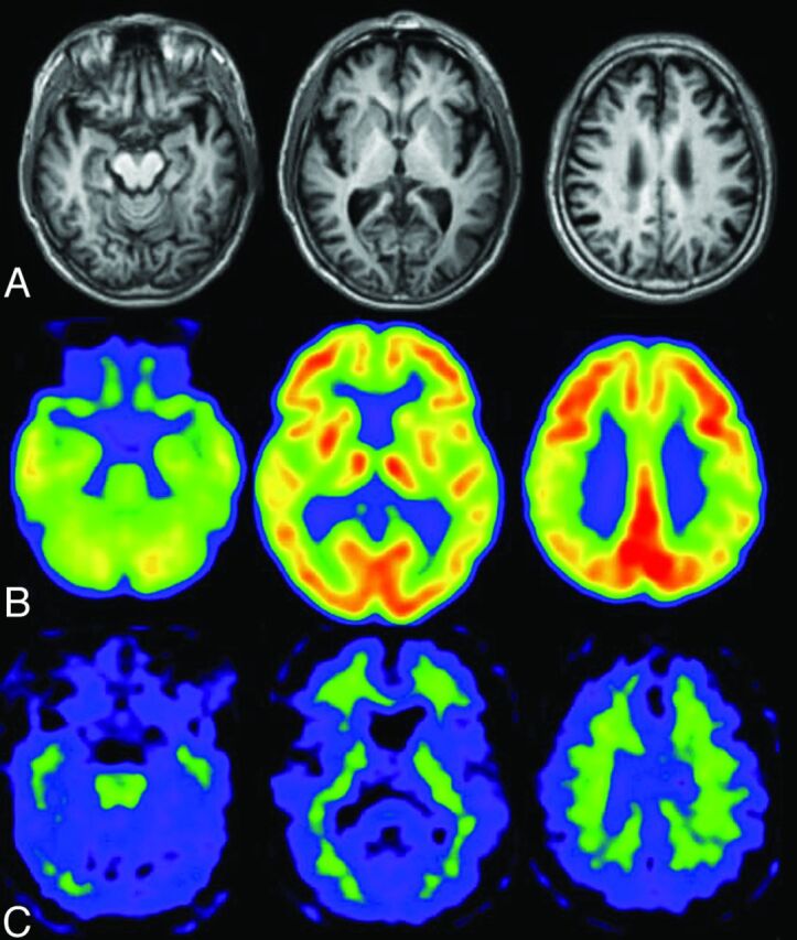
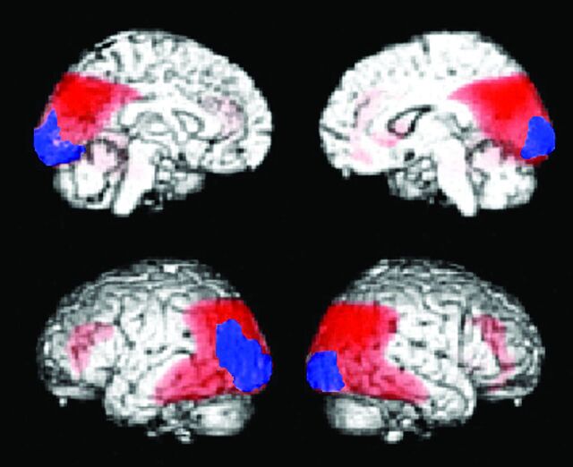
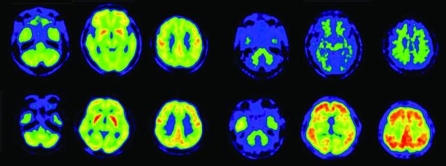
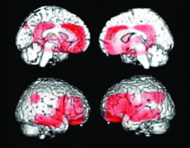
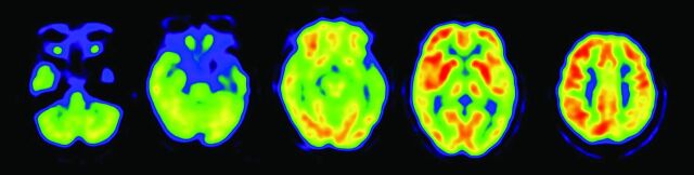
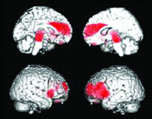
There is increasing use of neuroimaging modalities, including PET, for diagnosing dementia. For example, FDG-PET demonstrates hypometabolic regions in the posterior cingulate gyri, precuneus, and parietotemporal association cortices, while amyloid PET indicates amyloid deposition in Alzheimer disease and mild cognitive impairment due to Alzheimer disease. Furthermore, the use of combination PET with structural MR imaging can improve the diagnostic accuracy of dementia. In other neurodegenerative dementias, each disease exhibits a specific metabolic reduction pattern. In dementia with Lewy bodies, occipital glucose metabolism is decreased, while in frontotemporal dementia, frontal and anterior temporal metabolism is predominantly decreased. These FDG-PET findings and positive or negative amyloid deposits are important biomarkers for various neurodegenerative dementias.


Figures

Fig 1.
Fig 1.
A patient with early Alzheimer disease, 77 years of age, Mini-Mental State Examination score = 25. A, Minimal atrophy was seen in the right hippocampus. B, FDG-PET shows reduced glucose metabolism in the bilateral parietotemporal association cortices and posterior cingulate gyri and precuneus. C, PiB accumulations are demonstrated in the cerebral cortices except for the occipital and medial temporal regions. Medial parietal and frontal accumulations of PiB are high, indicative of positive amyloid deposit.
Fig 2.
Fig 2.
Healthy elderly male subject, 78 years of age, Mini-Mental State Examination score = 30. A, A slight enlargement of the right inferior horn of the lateral ventricle is seen on the T1-weighted MR image. B, The regional glucose metabolism is not reduced on the FDG-PET images. Note that the posterior cingulate glucose metabolism is much larger than that in other regions. C, PiB-PET shows nonspecific accumulation in the white matter but no PiB accumulation in the gray matter. The amyloid deposit is negative.
Fig 3.
Fig 3.
MCI due to AD. Regions exhibiting a significant reduction in glucose metabolism in patients with MCI due to AD (n = 20) compared with healthy elderly subjects (n = 20) are demonstrated by statistical parametric maps. Bilateral parietal and posterior cingulate metabolism is decreased in patients with MCI due to AD. These decreased regions are the same as those in patients with early AD.
Fig 4.
Fig 4.
Process of voxel-based statistical maps. Original FDG-PET image of a patient with mild AD (A) is analyzed by using the 3D-SSP program, and the 3D-SSP surface map and z score map are produced. The z score map shows the regions with significantly reduced metabolism compared with a normal data base (B), which aid in the diagnosis of AD.
Fig 5.
Fig 5.
Decreased glucose metabolic regions of DLB (n = 20). Statistical parametric maps show the areas where glucose metabolism is significantly decreased compared with age-matched healthy controls (n = 20). The red area indicates the parietotemporal association area, and the posterior cingulate gyri overlap the area where glucose metabolism is decreased in AD. The blue area indicates occipital cortices that are specific for DLB, where the glucose metabolism is preserved in AD (n = 20).
Fig 6.
Fig 6.
PiB-PET and FDG-PET images of patients with DLB. Upper row shows a 76-year-old man with DLB. This patient has Parkinsonism and cognitive fluctuation. His Mini-Mental State Examination score was 19. Diffuse glucose metabolic reduction is demonstrated in all regions apart from the striatum and primary sensorimotor cortices (upper left). PiB-PET (upper right) demonstrates a negative amyloid deposit. Lower row shows a 77-year-old woman with DLB, Parkinsonism, and cognitive fluctuation. Her Mini-Mental State Examination score was 23. FDG-PET demonstrates decreased glucose metabolism in the parietotemporal and frontal association cortices, occipital cortices, and posterior cingulate gyri, while metabolism in the striatum and primary sensorimotor metabolism is spared (lower left). PiB-PET demonstrated diffuse amyloid deposition in the cerebral cortices (lower right).
Fig 7.
Fig 7.
Hypometabolic regions in FTD. Statistical parametric mapping analysis shows hypometabolic regions in patients with FTD (n = 14) compared with healthy age-matched subjects (n = 20). Glucose metabolism in subjects with FTD was significantly decreased in the bilateral lateral and medial frontal cortices, the posterior cingulate gyrus, and small regions in the parietal association cortices.
Fig 8.
Fig 8.
FDG-PET image of a 54-year-old female patient with semantic dementia. A marked decreased left anterior temporal metabolism is shown on the FDG-PET image.
Fig 9.
Fig 9.
Hypometabolic regions in PSP. Statistical parametric mapping analysis shows hypometabolic regions in patients with PSP (n = 16) compared with normal age-matched subjects (n = 20). Glucose metabolism in subjects with PSP is significantly decreased in the bilateral lateral and medial frontal cortices and midbrain. The significant decreased midbrain metabolism is a hallmark of PSP.

References

    1. Ishii K. Clinical application of positron emission tomography for diagnosis of dementia. Ann Nucl Med 2002;16:515–25 - PubMed
    1. McKhann GM, Knopman DS, Chertkow H, et al. . The diagnosis of dementia due to Alzheimer's disease: recommendations from the National Institute on Aging-Alzheimer's Association workgroups on diagnostic guidelines for Alzheimer's disease. Alzheimers Dement 2011;7:263–69 - PMC - PubMed
    1. Albert MS, DeKosky ST, Dickson D, et al. . The diagnosis of mild cognitive impairment due to Alzheimer's disease: recommendations from the National Institute on Aging-Alzheimer's Association workgroups on diagnostic guidelines for Alzheimer's disease. Alzheimers Dement 2011;7:270–79 - PMC - PubMed
    1. McKhann G, Drachman D, Folstein M, et al. . Clinical diagnosis of Alzheimer's disease: report of the NINCDS-ADRDA Work Group under the auspices of Department of Health and Human Services Task Force on Alzheimer's Disease. Neurology 1984;34:939–44 - PubMed
    1. Ishii K, Sasaki M, Yamaji S, et al. . Relatively preserved hippocampal glucose metabolism in mild Alzheimer's disease. Dement Geriatr Cogn Disord 1998;9:317–22 - PubMed

Publication types

Substances

LinkOut - more resources