. 2013 Sep;123(9):4010-22.
doi: 10.1172/JCI69443. Epub 2013 Aug 15.

Schnurri-3 regulates ERK downstream of WNT signaling in osteoblasts

Affiliations

Schnurri-3 regulates ERK downstream of WNT signaling in osteoblasts

Jae-Hyuck Shim et al. J Clin Invest. 2013 Sep.

Abstract

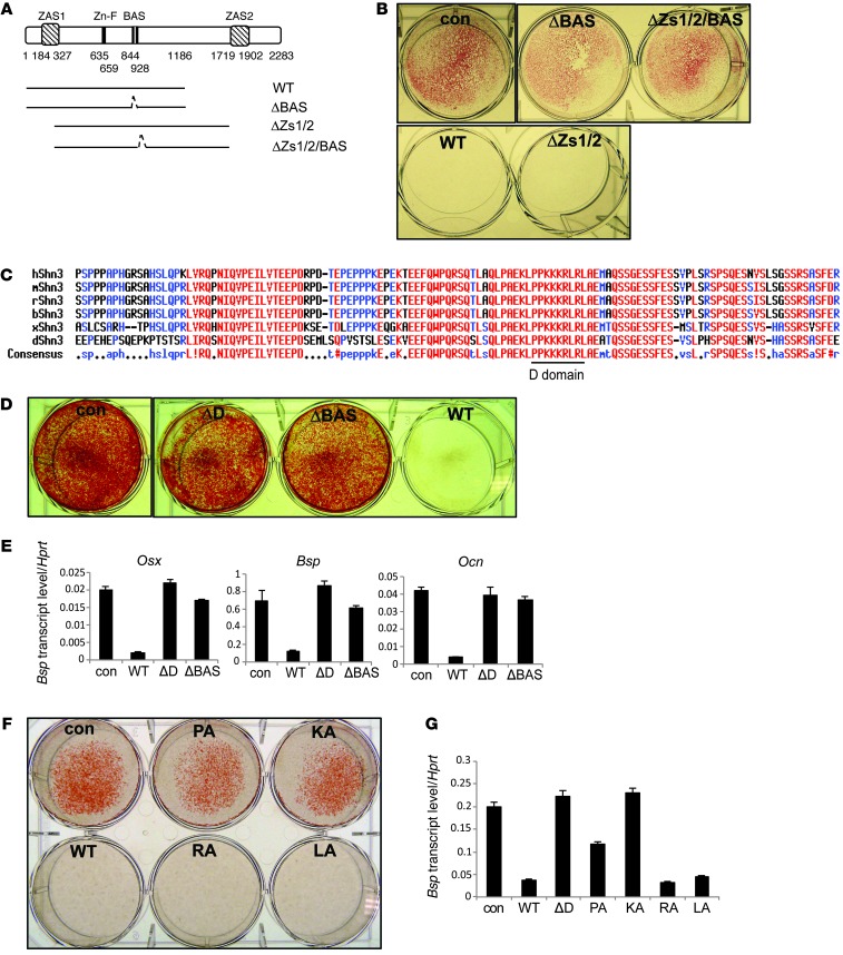
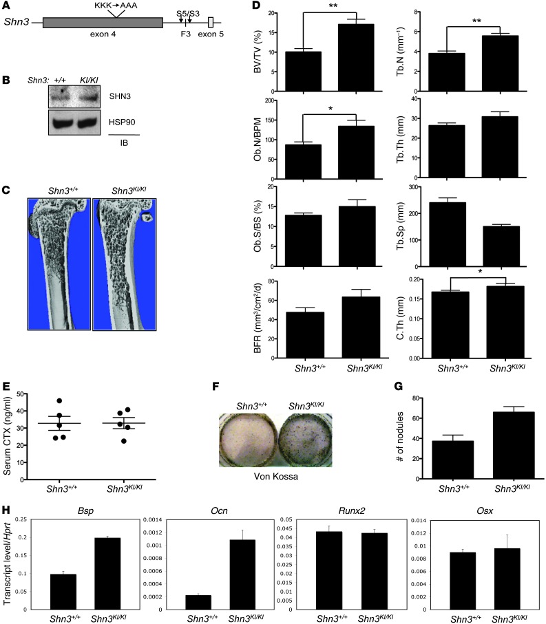
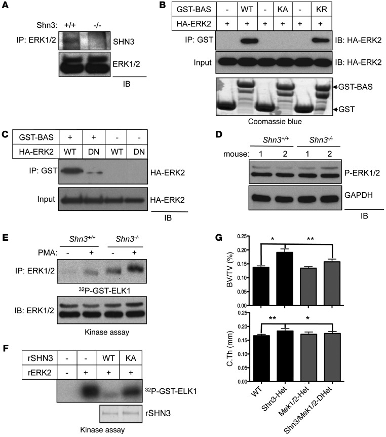
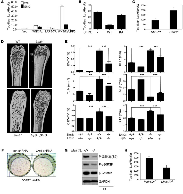
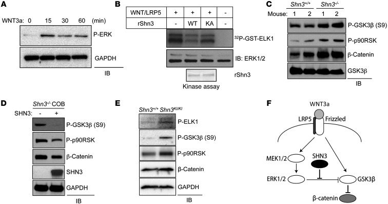
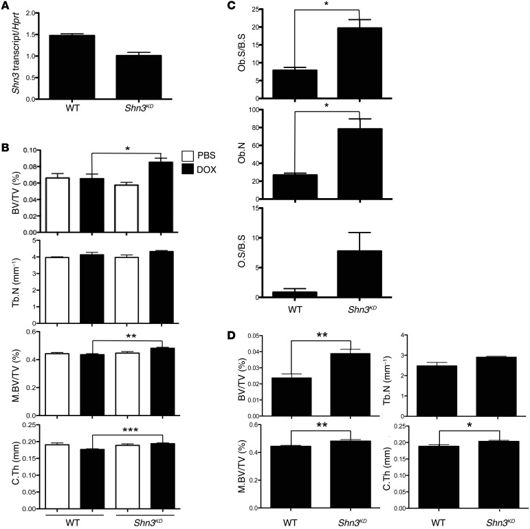
Mice deficient in Schnurri-3 (SHN3; also known as HIVEP3) display increased bone formation, but harnessing this observation for therapeutic benefit requires an improved understanding of how SHN3 functions in osteoblasts. Here we identified SHN3 as a dampener of ERK activity that functions in part downstream of WNT signaling in osteoblasts. A D-domain motif within SHN3 mediated the interaction with and inhibition of ERK activity and osteoblast differentiation, and knockin of a mutation in Shn3 that abolishes this interaction resulted in aberrant ERK activation and consequent osteoblast hyperactivity in vivo. Additionally, in vivo genetic interaction studies demonstrated that crossing to Lrp5(-/-) mice partially rescued the osteosclerotic phenotype of Shn3(-/-) mice; mechanistically, this corresponded to the ability of SHN3 to inhibit ERK-mediated suppression of GSK3β. Inducible knockdown of Shn3 in adult mice resulted in a high-bone mass phenotype, providing evidence that transient blockade of these pathways in adults holds promise as a therapy for osteoporosis.


Figures

Figure 1
Figure 1. Screening for functional motifs in SHN3.
All aa numbering is relative to the reference sequence NP_001121186.1. (A) Truncated SHN3 mutants (1–2,283 aa). WT, 1–1,186 aa; ΔBAS, Δ844–928 aa in 1–1,186 aa; ΔZs1/2 (ZAS1 and ZAS2 deletion), 327–1,719 aa; ΔZs1/2/BAS (ZAS1, ZAS2, and BAS deletion), Δ844–928 in 327–1,719 aa. (B, D, and F) hMSCs were infected with control lentivirus or lentiviruses encoding the indicated SHN3 constructs and cultured in differentiation medium for 21 days. Mineralization activity was analyzed by alizarin red staining. ΔD, D-domain deletion. (C) The aa sequence of the critical region of the BAS domain (p.810–933 aa), containing the D-domain (p.902–910 aa). (E and G) RNA levels of Osx, Bsp, and Ocn, analyzed by RT-PCR, in hMSCs infected by lentiviruses expressing the indicated SHN3 constructs or a control. Values in G are normalized to Hprt. Results are presented as mean + SD.
Figure 2
Figure 2. In vivo function of the 3-lysine motif of SHN3 in bone formation.
(A) The 3 lysine-to-alanine alleles knocked in to the endogenous Shn3 locus. Arrowheads denote position of primers (S5 and S3) used for PCR genotyping. (B) Primary Shn3+/+ and Shn3KI/KI BMSCs were lysed and immunoblotted with anti-SHN3 antibody. (C) μCT of 8-week-old Shn3+/+ and Shn3KI/KI mouse proximal femurs. (D) Histomorphometric and μCT analysis of 8-week-old Shn3+/+ and Shn3KI/KI mouse tibias. BV/TV, bone volume fraction; Ob.N/BPM, osteoblast number per bone perimeter; Ob.S/BS, osteoblast surface relative to bone surface; BFR, bone formation rate; Tb.N, trabecular number; Tb.Th, trabecular thickness; Tb.Sp, trabecular space; C.Th, cortical thickness. (E) Serum CTX levels in 8-week-old Shn3+/+ and Shn3KI/KI mice were determined by ELISA. (FH) Shn3+/+ and Shn3KI/KI BMSCs were cultured in differentiation medium, and mineralization was analyzed by Von Kossa staining (F) and quantization of mineralized nodules (G). Alternatively, total RNA was extracted for RT-PCR analysis (H), and values normalized to Hprt. Results are presented as mean + SD. *P < 0.05, **P < 0.005, Student’s t test.
Figure 3
Figure 3. SHN3 inhibits ERK MAPK activity in osteoblasts via the D-domain 3 lysine motif.
(A) Immortalized Shn3+/+ and Shn3–/– COBs were lysed, immunoprecipitation was performed with anti-ERK1/2 antibody and protein A–agarose, and the resulting complex was immunoblotted with an anti-SHN3 antibody. (B and C) HA-ERK2 (WT or DN [D319N]) was incubated with recombinant GST or with GST-tagged BAS, KA-BAS, or KR-BAS. GST-containing proteins were immunoprecipitated with glutathione-agarose, and the resulting complex was immunoblotted with anti-HA antibody. Input indicates loading controls of HA-ERK2, GST, or GST-BAS protein. (D) Primary COBs were isolated from Shn3+/+ and Shn3–/– mice, lysed, and immunoblotted with anti–phospho-ERK1/2 antibody. GAPDH was used as a cytosolic protein control. (E) Immortalized Shn3+/+ and Shn3–/– COBs were stimulated with PMA for 30 minutes, and then ERK1/2 was immunoprecipitated. The immunoprecipitates were mixed with recombinant ELK1, and ERK kinase activity was analyzed by in vitro kinase assay. (F) Recombinant ERK2 (rERK2) was incubated with or without the indicated recombinant His-SHN3 (rSHN3) along with GST-ELK1, and ERK kinase activity was analyzed by in vitro kinase assay. Coomassie blue staining indicates loading control of recombinant His-SHN3. (G) μCT analysis of 5-week-old WT (Shn3fl/+), Shn3-Het, Mek1/2-Het, and Shn3/Mek1/2-DHet mouse femurs. Bone volume fraction and cortical thickness are shown. Results are presented as mean + SD. *P < 0.05, **P < 0.005, Student’s t test.
Figure 4
Figure 4. SHN3 inhibits the WNT-mediated β-catenin pathway via ERK regulation.
(A and B) C3H10T1/2 cells were infected by lentivirus expressing vector or SHN3-WT (A) or SHN3-KA (B) and transfected with top-flash luc and Renilla along with a xWNT8/Fz5 fusion protein, a constitutively active mutant of LRP5 (LRP5-CA), or xWNT8/Fz5 fusion protein plus LRP5. Relative luciferase activity normalized to Renilla is shown. (C) Shn3+/+ and Shn3–/– COBs were transfected with top-flash luc along with Renilla. Luciferase activity was analyzed 6 days after culture in differentiation medium. Values are normalized to Renilla. (D and E) μCT analysis of 8-week-old Shn3+/+, Lrp5–/–, Shn3–/–, and Lrp5–/–;Shn3–/– mouse femurs. (D) 3-dimensional reconstructions of proximal femur. (E) Bone volume fraction, trabecular number, trabecular thickness, cortical bone volume fraction (C.BV/TV), trabecular space, and cortical thickness. (F) Primary Shn3–/– COBs were infected by control or Lrp5 shRNA–expressing lentivirus and cultured in differentiation medium, and mineralization activity was analyzed by alizarin red staining. (G) Primary Mek1fl/fl;Mek2–/– COBs were infected by lentivirus expressing vector (Mek1/2+/+) or Cre recombinase (Mek1/2–/–), cultured in differentiation medium, lysed, and immunoblotted with the indicated antibodies. (H) Primary Mek1fl/fl;Mek2–/– COBs infected as in G were transfected with top-flash luc and Renilla. Luciferase activity was analyzed 6 days after culture in differentiation medium. Values are normalized to Renilla. Results are presented as mean + SD. **P < 0.005, ***P < 0.0005, Student’s t test.
Figure 5
Figure 5. SHN3 regulates GSK3β activity and β-catenin level in osteoblasts.
(A) Primary WT COBs were stimulated with murine WNT3a (200 ng/ml) for the indicated time points after 12 hour serum starvation. Cells were lysed and immunoblotted with antibodies specific to phospho-ERK and GAPDH. (B) HEK293 cells were transfected with a xWNT8/Fz5 fusion protein along with LRP5, lysed, and immunoprecipitated by anti-ERK1/2 antibody and protein A–agarose. The immunoprecipitates were incubated with GST-ELK1 and either His-SHN3-WT or His-SHN3-KA, and ERK kinase activity was analyzed by in vitro kinase assay. Inputs indicate loading control of recombinant His-SHN3. (C and E) Primary Shn3+/+ and Shn3–/– (C) or Shn3KI/KI (E) COBs were cultured in differentiation medium, lysed, and immunoblotted with the indicated antibodies. (D) Primary Shn3–/– COBs were infected with lentivirus expressing vector or SHN3 and cultured in differentiation medium containing puromycin for 6 days. Cells were lysed and immunoblotted with the indicated antibodies. (F) SHN3 regulation of ERK activity in the context of WNT/β-catenin signaling.
Figure 6
Figure 6. Inducible knockdown of SHN3 increases bone mass in adult mice.
(A) 8-week-old WT and Shn3KD mice were fed DOX-containing chow for 6 weeks, and total RNA was extracted from long bones for RT-PCR analysis. Values are normalized to Hprt. (B) 8-week-old WT and Shn3KD mice were fed PBS- or DOX-containing chow for 6 weeks. Femurs from 14-week-old mice were analyzed by μCT. Shown are bone volume fraction, trabecular number, midshaft bone volume fraction (M.BV/TV), and cortical thickness. (C) 8-week-old WT and Shn3KD mice were fed DOX-containing chow for 6 weeks, and femurs from 14-week-old mice were analyzed by histomorphometric analysis. (D) 18-week-old WT and Shn3KD mice were fed DOX-containing chow for 6 weeks, and femurs from 24-week-old mice were analyzed by μCT. Results are presented as mean + SD. *P < 0.05, **P < 0.005, ***P < 0.0005, Student’s t test.

References

    1. Hill TP, Spater D, Taketo MM, Birchmeier W, Hartmann C. Canonical Wnt/β-catenin signaling prevents osteoblasts from differentiating into chondrocytes. Dev Cell. 2005;8(5):727–738. doi: 10.1016/j.devcel.2005.02.013. - DOI - PubMed
    1. Day TF, Guo X, Garrett-Beal L, Yang Y. Wnt/β-catenin signaling in mesenchymal progenitors controls osteoblast and chondrocyte differentiation during vertebrate skeletogenesis. Dev Cell. 2005;8(5):739–750. doi: 10.1016/j.devcel.2005.03.016. - DOI - PubMed
    1. Kolpakova E, Olsen BR. Wnt/β-catenin — a canonical tale of cell-fate choice in the vertebrate skeleton. Dev Cell. 2005;8(5):626–627. doi: 10.1016/j.devcel.2005.04.008. - DOI - PubMed
    1. Clevers H, Nusse R. Wnt/β-catenin signaling and disease. Cell. 2012;149(6):1192–1205. doi: 10.1016/j.cell.2012.05.012. - DOI - PubMed
    1. MacDonald BT, He X. Frizzled and LRP5/6 receptors for Wnt/β-catenin signaling. Cold Spring Harb Perspect Biol. 2012. 4 (12).pii: a007880 10.1101/cshperspect.a007880 - DOI - PMC - PubMed

Publication types

MeSH terms