. 2013 Sep;4(9):1373-87.
doi: 10.18632/oncotarget.1165.

Crucial role for early growth response-1 in the transcriptional regulation of miR-20b in breast cancer

Affiliations

Crucial role for early growth response-1 in the transcriptional regulation of miR-20b in breast cancer

Dongping Li et al. Oncotarget. 2013 Sep.

Abstract

Transcriptional regulation of miRNAs that control the pathogenesis of breast cancer remains largely unknown. Here, we showed that ionizing radiation, a known breast carcinogen, triggered the differential expression of miR-20b in mammary tissues. We identified several GC-rich consensus binding motifs for the zinc finger transcription factor early growth response-1 (EGR1) in miR-20b promoter. miR-20b was upregulated by IR and its upregulation correlated with EGR1 expression in the breast cancer cell line HCC1806. Therefore, we used HCC1806 cells as a model system to explore the role of EGR1 in miR-20b transcription. siRNA knockdown of EGR1 attenuated miR-20b expression. Luciferase assays showed that whereas EGR1 stimulated luciferase activity driven by the wild-type miR-20b promoter, this induction was abolished in the mutant miR-20 promoter construct. We noted significant enrichment of EGR1 at miR-20b promoter in HCC1806 cells compared with normal human mammary epithelial cells. Suppression of miR-20b significantly inhibited HCC1806 cell proliferation and migration, and led to G0/G1 and S phase arrest. In vitro RNA-pull down assays indicated that miR-20b targets numerous tumor suppressors, including PTEN and BRCA1, which were downregulated in HCC1806. Conversely, suppression of miR-20b increased PTEN and BRCA1 levels. Moreover, immunohistochemical and FISH analyses showed that the miR-20b expression correlated significantly with EGR1 levels in breast cancer tissues. Our findings thus demonstrate for the first time that EGR1 is a key player in the transcriptional control of miR-20b, and miR-20b may in turn function as an oncogene by contributing to breast tumorigenesis via tumor suppressor targeting.


Figures

Figure 1
Figure 1. IR induces miR-20b expression in mammary gland tissues/cells in a dose- and time-dependent manner
(A and B) Total RNA isolated from the mammary gland tissues of six-week-old female Long Evans rats exposed to either 30 kVp/0.1 Gy, 80 kVp/2.5 Gy X-ray, or sham-treatment 96 hours post-irradiation was subjected to microRNA microarray; the levels of rno-miR-20b were determined by real-time RT-PCR. (C) Total RNA was isolated from the mammary gland tissues of six-week-old female Long Evans rats at different time points post-IR, and the levels of rno-miR-20b were examined by real-time RT-PCR. (D and E) Total RNA isolated from HMEC exposed to either 30 kVp/0.1 Gy or 80 kVp/2.5 Gy X-ray was subjected to real-time RT-PCR using primers for hsa-miR-20b and EGR1. The hash indicates p<0.1; the asterisk indicates p<0.05.
Figure 2
Figure 2. EGR1 correlates with miR-20b expression levels
(A) Total RNA isolated from HMEC and breast cancer cell lines MCF7, ZR75-1, HCC1419, and HCC1806 was subjected to real-time RT-PCR with a primer set for miR-20b. (B) Whole cell lysates prepared from the above cell lines were subjected to Western blot analysis using antibodies against EGR1 and GAPDH. (C) HCC1806 cells were transiently transfected with either siEGR1 (siRNA targeting EGR1) or control siRNA; the levels of EGR1 mRNA and protein were determined by real-time RT-PCR (upper panel) and Western blot analysis (lower panel). (D) HCC1806 cells were transiently transfected with either siEGR1 or control siRNA; the levels of miR-20b were determined by real-time RT-PCR. The asterisk indicates p<0.05.
Figure 3
Figure 3. EGR1 regulates miR-20b transcription
(A) The wild-type and mutant miR-20b promoter reporters used in this project. (B) HEK293 cells were transiently transfected with pGL3-WT-miR20b-Prom or pGL3-MT-miR20b-Prom and pCB6-Egr1 or pCB6; luciferase activity was detected according to the manufacturer's instruction. (C) Real-time ChIP-PCR and conventional ChIP-PCR were performed as described in “Materials and Methods”. (D) Nuclear extracts were prepared from HCC1806 cells, and EMSA was performed using ChIP-grade antibody to EGR1 according to the manufacturer's instruction. The asterisk indicates p<0.05.
Figure 4
Figure 4. miR-20b inhibitor suppresses breast cancer cell proliferation and migration, as well as induces G0/G1 and S phase arrest
(A) HCC1806 cells were transfected with either miR-20b inhibitor or a negative control; MTT assay (cell proliferation assay) was performed according to the manufacturer's instruction. (B and C) HCC1806 cells were transfected with either miR-20b inhibitor or negative control; 24 hours after transfection, wound-healing assay and statistical analysis of migrated cells were performed. (D) HCC1806 cells were transfected with either miR-20b inhibitor or negative control; 96 hours after transfection, cell cycle analysis was conducted using DB FACSCanto II Flow Cytometer. The asterisk indicates p<0.05.
Figure 5
Figure 5. PTEN and BRCA1 are direct targets of miR-20b
(A) The network of the predicted targets of hsa-miR-20b was generated using STRING 9.0. (B) Diagram of 3'UTR sequences of PTEN and BRCA1 targeted by hsa-miR-20b. (C) Whole cellular lysates prepared from HMEC, HCC1806, and HCC1806 transfected with either 50 nM miR-20b inhibitor or non-specific control for 72 hours were subjected to Western blot analysis using antibodies specific to PTEN and BRCA1. (D) HEK293 cells grown to 90% confluency were cotransfected with either pGL3-PTEN or pGL3-BRCA1 reporter, and the indicated concentration of hsa-miR-20b or 50 nM nonspecific miRNA as a control; 24 hours after transfection, luciferase activity was detected using Dual-Luciferase Reporter Assay System according to the manufacturer's instruction. The asterisk indicates p<0.05.
Figure 6
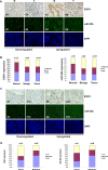
Figure 6. EGR1 expression is correlated with miR-20b expression in breast cancer tissues
(A) Representatives of EGR1 and hsa-miR-20b stainings in the same sections of breast cancer tissue arrays. (B) Statistical and correlation analyses of EGR1 and hsa-miR-20b expression in breast cancer tissues. (C) Representatives of EGR1 and hsa-miR-20b staining in the same sections of metastatic breast cancer tissue arrays. (D) Statistical and correlation analyses of EGR1 and hsa-miR-20b expression in metastatic breast cancer tissues.
Figure 7
Figure 7. miR-20b transcriptionally activated by EGR1 directly targets PTEN and BRCA1 in breast cancer
Serum-inducible zinc finger transcription factor EGR1 is induced and activated in response to a wide range of extracellular stimuli, including growth factors, cytokines, UV light, ionizing radiation, and mechanical injury. Once activated, EGR1 translocates into nucleus and binds to the consensus motifs at miR-20b promoter, leading to miR-20b transcription. The mature miR-20b assembles with other proteins to form RNA-induced silencing complex (RISC), the later recognizes and binds to PTEN and BRCA1 mRNAs, leading to either translational suppression or degradation of those two molecules, consequently resulting in breast cancer cell proliferation and migration.

References

    1. Schairer C, Mink PJ, Carroll L, Devesa SS. Probabilities of death from breast cancer and other causes among female breast cancer patients. J Natl Cancer Inst. 2004;96:1311–21. - PubMed
    1. Mattsson A, Rudén BI, Hall P, Wilking N, Rutqvist LE. Radiation-induced breast cancer: Long-term follow-up of radiation therapy for benign breast disease. J Natl Cancer Inst. 1993;85:1679–85. - PubMed
    1. Doody MM, Lonstein JE, Stovall M, Hacker DG, Luckyanov N, Land CE. Breast cancer mortality after diagnostic radiography: findings from U.S. Scoliosis Cohort Study. Spine (Phila Pa 1976) 2000;25:2052–63. - PubMed
    1. Ronckers CM, Erdmann CA, Land CE. Radiation and breast cancer: a review of current evidence. Breast Cancer Res. 2005;7:21–32. - PMC - PubMed
    1. Stecklein SR, Jensen RA, Pal A. Genetic and epigenetic signatures of breast cancer subtypes. Front Biosci (Elite Ed) 2012;4:934–49. - PubMed

Publication types

MeSH terms