

microRNAs derived from circulating exosomes as noninvasive biomarkers for screening and diagnosing lung cancer
- PMID: 23945385
- PMCID: PMC4123222
- DOI: 10.1097/JTO.0b013e318299ac32
microRNAs derived from circulating exosomes as noninvasive biomarkers for screening and diagnosing lung cancer
Abstract
Introduction: Lung cancer is the highest cause of mortality among tumor pathologies worldwide. There are no validated techniques for an early detection of pulmonary cancer lesions other than low-dose helical computed tomography scan. Unfortunately, this method has some negative effects. Recent studies have laid the basis for development of exosomes-based techniques to screen/diagnose lung cancers. As the isolation of circulating exosomes is a minimally invasive procedure, this technique opens new possibilities for diagnostic applications.
Methods: We used a first set of 30 plasma samples from as many patients, including 10 patients affected by lung adenocarcinomas, 10 with lung granulomas, and 10 healthy smokers matched for age and sex as negative controls. Wide-range microRNAs analysis (742 microRNAs) was performed by quantitative real time polymerase chain reaction. Data were compared on the basis of lesion characteristics, using WEKA software for statistics and modeling. Subsequently, selected microRNAs were evaluated on an independent larger group of samples (105 specimens: 50 lung adenocarcinomas, 30 lung granulomas, and 25 healthy smokers).
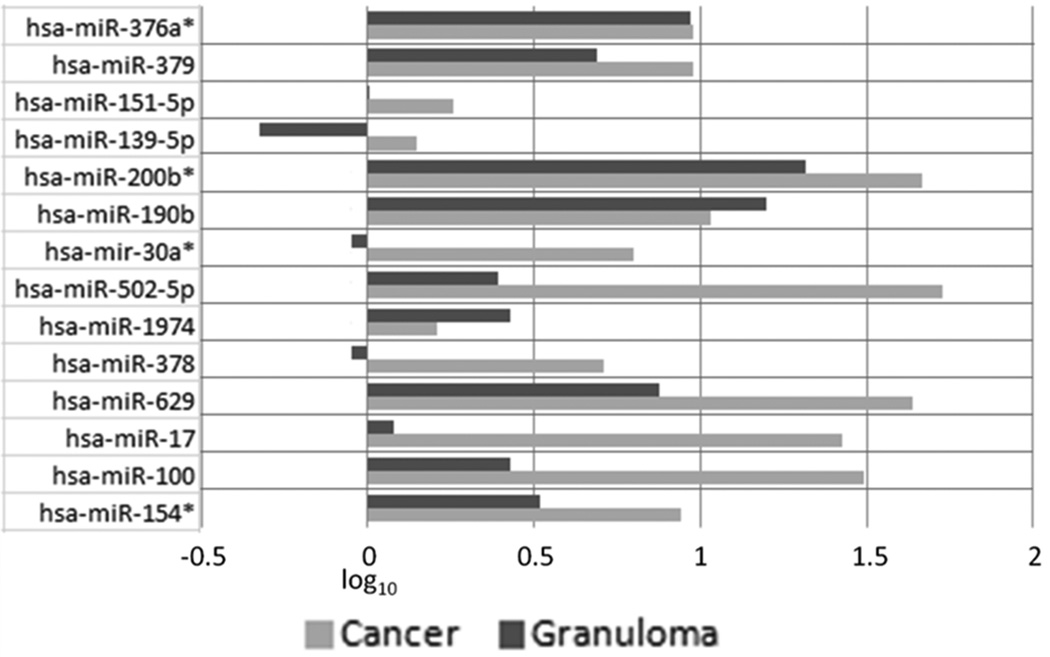
Results: This analysis led to the selection of four microRNAs to perform a screening test (miR-378a, miR-379, miR-139-5p, and miR-200b-5p), useful to divide population into two groups: nodule (lung adenocarcinomas + carcinomas) and non-nodule (healthy former smokers). Six microRNAs (miR-151a-5p, miR-30a-3p, miR-200b-5p, miR-629, miR-100, and miR-154-3p) were selected for a second test on the nodule population to discriminate between lung adenocarcinoma and granuloma.
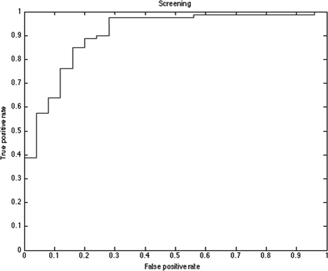
Conclusions: The screening test showed 97.5% sensitivity, 72.0% specificity, and area under the curve receiver operating characteristic of 90.8%. The diagnostic test had 96.0% sensitivity, 60.0% specificity, and area under the curve receiver operating characteristic of 76.0%. Further evaluation is needed to confirm the predictive power of these models on larger cohorts of samples.
Figures

References
-
- Siegel R, Ward E, Brawley O, et al. Cancer statistics 2011, The impact of eliminating socioeconomic and racial disparities on premature cancer deaths. CA Cancer J Clin. 2011;61:212–236. - PubMed
-
- Cortez MA, Calin GA. MicroRNA identification in plasma and serum: a new tool to diagnose and monitor diseases. Expert Opin Biol Ther. 2009;9:703–711. - PubMed
Publication types
MeSH terms
Substances
Grants and funding
LinkOut - more resources
Full Text Sources
Other Literature Sources
Medical
Research Materials
