

Comment
doi: 10.1038/nature12549.
Epub 2013 Aug 14.
Metabolism: Sweet enticements to move
- PMID: 23945589
- PMCID: PMC4554390
- DOI: 10.1038/nature12549
Item in Clipboard
Comment
Metabolism: Sweet enticements to move
Nature.
.
Abstract
The formation of new blood vessels from pre-existing ones is a carefully orchestrated dance. A study reveals that the metabolism of sugar by glycolysis contributes to its regulation.
Figures
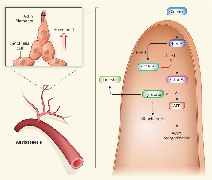
The formation of new blood vessels involves the outward movement of endothelial
cells from the lining of existing blood vessels, a process that relies on the
rapid reorganization of actin-protein filaments in cellular structures called
filopodia and lamellipodia (not shown). The energy for this (in the form of ATP)
is provided by the breakdown of glucose, but endothelial cells are unusual in
that the pyruvate produced by glycolysis is converted to lactate, rather than
being channelled into mitochondria for further oxidation, as occurs in most
cells. De Bock et al. show that both angiogenesis and glycolysis are
accelerated by the activity of the enzyme PFK2 in endothelial-cell lamellipodia
and filopodia. PFK2 converts the glycolytic intermediate fructose-6-phosphate
(F-6-P) into fructose-2,6-bisphosphate (F-2,6-P2), which, in turn,
enhances the activity of the glycolytic enzyme PFK1, thereby accelerating
glycolysis at these sites. Pyruvate then leaves the cell as lactate, probably
because filopodia and lamellipodia are too small to accommodate
mitochondria.
Comment on
-
Role of PFKFB3-driven glycolysis in vessel sprouting.Cell. 2013 Aug 1;154(3):651-63. doi: 10.1016/j.cell.2013.06.037. Cell. 2013. PMID: 23911327
References
Publication types
MeSH terms
Substances
Grants and funding
LinkOut - more resources
Full Text Sources
Other Literature Sources
Miscellaneous
