. 2013 Nov;41(20):9484-99.
doi: 10.1093/nar/gkt720. Epub 2013 Aug 14.

Functional assignment of KEOPS/EKC complex subunits in the biosynthesis of the universal t6A tRNA modification

Affiliations

Functional assignment of KEOPS/EKC complex subunits in the biosynthesis of the universal t6A tRNA modification

Ludovic Perrochia et al. Nucleic Acids Res. 2013 Nov.

Abstract

N(6)-threonylcarbamoyladenosine (t(6)A) is a universal tRNA modification essential for normal cell growth and accurate translation. In Archaea and Eukarya, the universal protein Sua5 and the conserved KEOPS/EKC complex together catalyze t(6)A biosynthesis. The KEOPS/EKC complex is composed of Kae1, a universal metalloprotein belonging to the ASHKA superfamily of ATPases; Bud32, an atypical protein kinase and two small proteins, Cgi121 and Pcc1. In this study, we investigated the requirement and functional role of KEOPS/EKC subunits for biosynthesis of t(6)A. We demonstrated that Pcc1, Kae1 and Bud32 form a minimal functional unit, whereas Cgi121 acts as an allosteric regulator. We confirmed that Pcc1 promotes dimerization of the KEOPS/EKC complex and uncovered that together with Kae1, it forms the tRNA binding core of the complex. Kae1 binds l-threonyl-carbamoyl-AMP intermediate in a metal-dependent fashion and transfers the l-threonyl-carbamoyl moiety to substrate tRNA. Surprisingly, we found that Bud32 is regulated by Kae1 and does not function as a protein kinase but as a P-loop ATPase possibly involved in tRNA dissociation. Overall, our data support a mechanistic model in which the final step in the biosynthesis of t(6)A relies on a strictly catalytic component, Kae1, and three partner proteins necessary for dimerization, tRNA binding and regulation.


Figures

Figure 1.
Figure 1.
Biosynthesis of t6A modification in Archaea and Eukarya. (A) Schematic representation of the anticodon loop of tRNALys from E. coli. N6-threonylcarbamoyladenosine (t6A) at the position 37 is indicated with full circle in dark gray and the major determinant nucleoside U36 is indicated in gray. Messenger RNA is indicated in black circles and the interaction of the anticodon with the codon triplet AAA is indicated by dotted lines. t6A stabilizes the anticodon loop structure by preventing intraloop bonding between U33 and A37. (B) Reaction scheme leading to t6A biosynthesis in Archaea and Eukarya. Sua5 catalyzes the formation of the l-threonyl-carbamoyl-AMP (TC-AMP) intermediate from bicarbonate, threonine and ATP. KEOPS catalyzes the final step of the reaction in which l-threonyl-carbamoyl moiety is transferred from TC-AMP to A37 of substrate tRNA. tRNA binding to KEOPS significantly stimulates hydrolysis of ATP by KEOPS. (C) Schematic representation of the architecture of the KEOPS complex. The arrangement of different subunits is represented according to the reported composite model structure (2). For Kae1, two domains of the ASHKA fold are indicated by I and II and the iron atom in the active site cavity is represented as a full circle.
Figure 2.
Figure 2.
In vitro biosynthesis of t6A-modified tRNA by subcomplexes of KEOPS from P. abyssi. Different subcomplexes and their combinations were incubated with Sua5 from P. abyssi, ATP, bicarbonate, C14threonine and substrate tRNA (see Materials and Methods). The radioactive l-threonine incorporated in TCA-precipitated material was counted by scintillation. Error bars correspond to standard deviation from three independent experiments. Single letter designations are as follows: P, Pcc1; K, Kae1; B, Bud32 and C, Cgi121.
Figure 3.
Figure 3.
ATPase activity of wild-type and mutant KEOPS proteins from P. abyssi. KEOPS subcomplexes were incubated with α-P32 ATP in reaction buffer either alone or in combination with other proteins. Produced radiolabeled (α-P32)-ADP was separated by TLC and visualized by phosphorimaging. When indicated, Ec_tRNALys produced in overexpressing E. coli strain was added to the reaction mixture. In negative controls, indicated by a minus sign, the proteins were omitted in the reaction mixture. (A): ATPase activity of binary complexes. (B): ATPase activity of ternary complex Kae1–Bud32–Cgi121. (C): The ATPase activity was measured for reconstituted KEOPS complexes containing point mutations in Kae1 and Bud32 indicated at the bottom. Single letter designations are as follows: P, Pcc1; K, Kae1; B, Bud32 and C, Cgi121.
Figure 4.
Figure 4.
Impact of point mutations of Kae1 and Bud32 on the in vitro biosynthesis of t6A modification by KEOPS from P. abyssi. Whole KEOPS complexes were reconstituted by mixing two binary subcomplexes carrying point mutations. The reconstituted complexes were incubated with Sua5 from P. abyssi, ATP, bicarbonate, C14 l-threonine and substrate tRNA. The reaction mixtures were precipitated with TCA, and the radioactivity levels in the precipitates were measured by scintillation counting. The counts were converted into amount of incorporated C14 l-threonine into tRNA using a standard calibration curve. The negative control, which is designated with a minus sign, corresponds to the total assay mixture without proteins.
Figure 5.
Figure 5.
Analysis of ATPase and protein kinase activity of Bud32. (A) As a part of the KEOPS complex, Bud32 does not exhibit a significant protein kinase activity. Sua5 and the KEOPS complex were incubated under standard t6A assay conditions in presence of γ-P32 ATP (see Materials and Methods). The reaction mixture was analyzed by SDS-PAGE (part a), and the radioactivity retained by the proteins was recorded by phosphorimaging (part b). The same reaction mixture (part c) and a negative control lacking proteins (part -) was analyzed by TLC. The radioactive spots correspond to different nucleotides and free inorganic phosphate, as indicated. (B) Bud32 exhibits autophosphorylation activity in presence of Cgi121. The Bud32–Cgi121 binary complex was incubated in presence of γ-P32 ATP under standard t6A assay conditions, except that tRNA was omitted in the assay. The reaction mixture was analyzed by SDS-PAGE (part a), and the radioactivity retained by the proteins was recorded by phosphorimaging (part b). An aliquot of the reaction mixture (part c) and a negative control lacking proteins (part -) was analyzed by TLC. (C) Bud32 phosphotransferase activity is switched off in presence of Kae1. The Bud32–Cgi121 complex was incubated in presence of γ-P32 ATP under standard t6A assay conditions, except that tRNA was omitted in the assay. Reaction mixtures contained Bud32–Cgi121 (BC) alone (leftmost sample) or BC mixed with increasing concentrations of Pcc1–Kae1 (PK). In the rightmost sample, PK and BC subcomplexes were present in the reaction mixture in equimolar amounts. The reaction mixtures were analyzed by SDS-PAGE, and the radioactivity retained by the proteins was recorded by phosphorimaging (upper panel). The production of free inorganic phosphate for each reaction mixture was monitored by TLC analysis (lower panel).
Figure 6.
Figure 6.
Molecular docking of TC-AMP molecule into the active site of Kae1 from P. abyssi. (A1) Active site cavity of Kae1 from P. abyssi with bound AMP-PNP (PDB file 2IVN). The nucleotide is shown as sticks and hydrogen bonds are indicated by dotted lines. (A2) Detailed view of the γ-phosphate binding site. (B1) Active site cavity of Kae1 from P. abyssi with docked TC-AMP. The thermodynamically most favorable conformation (ΔG = −9.7 kcal/mol) is presented. TC-AMP is shown as sticks and hydrogen bonds are indicated by dotted lines. The docking was performed as blind docking by using Autodock Vina 1.1.2 software (see ‘Materials and Methods’ section). B2. Detailed view of the threonyl binding site. Further details are described in the main text.
Figure 7.
Figure 7.
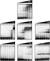
Binding of tRNA by PaKEOPS subcomplexes and individual proteins. Individual PaKEOPS subunits and subcomplexes were mixed with 10 nM of radiolabeled (P32) tRNA at room temperature and the mixture was separated by native PAGE (see ‘Materials and Methods’ section). The radioactive bands corresponding to unbound tRNA (at the bottom of each gel) and the nucleoprotein complexes were recorded by phosphorimaging. Protein concentrations used are indicated in µM at the top of each panel. In the downright panel, fixed concentration of the Kae1–Bud32 complex (0.5 µM) was titrated with increasing concentrations of Cgi121, which are indicated for each sample at the top of the gel. Minus sign stands for the control sample to which proteins were not added. Single letter designations are as follows: P, Pcc1; K, Kae1; B, Bud32 and C, Cgi121.
Figure 8.
Figure 8.
Putative mechanism for the catalysis of the last step in the biosynthesis of t6A modification by the KEOPS complex. (1) Formation of TC-AMP: Sua5 catalyzes the condensation of threonine, bicarbonate and ATP leading to the formation of an unstable TC-AMP intermediate and release of inorganic pyrophosphate. (2) Binding of tRNA and TC-AMP to KEOPS: TC-AMP binds into the active site of Kae1 and interacts directly with the iron atom via threonyl part of the molecule. Binding of tRNA to the complex provokes conformational changes in the complex, including the movement of the C-terminal tail of Bud32 (indicated with an arrow). Pcc1 and Kae1 are involved in the major part of contacts (gray triangles) with tRNA, whereas Bud32 participates in binding of tRNA via C-terminal tail. Anticodon loop carrying the target nucleotide A37 is positioned at the entrance to the active site cavity of Kae1 next to the TC-AMP intermediate and iron atom. (3) Transfer of l-threonyl-carbamoyl to tRNA: Threonyl-carbamoyl moiety is transferred to A37 of substrate tRNA in an ATP-independent fashion and AMP is released. (4) Release of t6A-modified tRNA: ATP hydrolysis catalyzed by Bud32 powers the conformational changes, in particular motion of the C-terminal tail of Bud32 (indicated by red arrow), which leads to the dissociation of modified tRNA from the KEOPS complex. The resulting KEOPS complex is competent for another catalytic cycle. Further details are given in the main text.

References

    1. Agris PF. Bringing order to translation: the contributions of transfer RNA anticodon-domain modifications. EMBO Rep. 2008;9:629–635. - PMC - PubMed
    1. Mao DYL, Neculai D, Downey M, Orlicky S, Haffani YZ, Ceccarelli DF, Ho JSL, Szilard RK, Zhang W, Ho CS, et al. Atomic Structure of the KEOPS Complex: An Ancient Protein Kinase-Containing Molecular Machine. Mol. Cell. 2008;32:259–275. - PMC - PubMed
    1. Ishikura H, Yamada Y, Murao K, Saneyoshi M, Nishimura S. The presence of N-[9-(beta-D-ribofuranosyl)purin-6-ylcarbamoyl]threonine in serine, methionine and lysine transfer RNA’s from Escherichia coli. Biochem. Biophys. Res. Commun. 1969;37:990–995. - PubMed
    1. Powers DM, Peterkofsky A. The presence of N-(purin-6-ylcarbamoyl)threonine in transfer ribonucleic acid species whose codons begin with adenine. J. Biol. Chem. 1972;247:6394–6401. - PubMed
    1. Grosjean H, Sprinzl M, Steinberg S. Posttranscriptionally modified nucleosides in transfer RNA: their locations and frequencies. Biochimie. 1995;77:139–141. - PubMed

Publication types

MeSH terms