

MicroRNA-31 predicts the presence of lymph node metastases and survival in patients with lung adenocarcinoma
- PMID: 23946296
- PMCID: PMC3823052
- DOI: 10.1158/1078-0432.CCR-13-0320
MicroRNA-31 predicts the presence of lymph node metastases and survival in patients with lung adenocarcinoma
Abstract
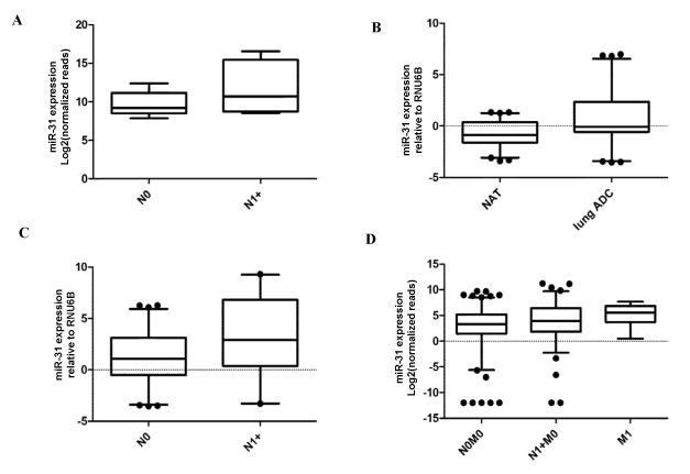
Purpose: We conducted genome-wide miRNA-sequencing (miRNA-seq) in primary cancer tissue from patients of lung adenocarcinoma to identify markers for the presence of lymph node metastasis.
Experimental design: Markers for lymph node metastasis identified by sequencing were validated in a separate cohort using quantitative PCR. After additional validation in the The Cancer Genome Atlas (TCGA) dataset, functional characterization studies were conducted in vitro.
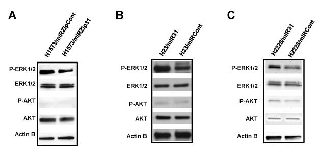
Results: MiR-31 was upregulated in lung adenocarcinoma tissues from patients with lymph node metastases compared with those without lymph node metastases. We confirmed miR-31 to be upregulated in lymph node-positive patients in a separate patient cohort (P = 0.009, t test), and to be expressed at higher levels in adenocarcinoma tissue than in matched normal adjacent lung tissues (P < 0.0001, paired t test). MiR-31 was then validated as a marker for lymph node metastasis in an external validation cohort of 233 lung adenocarcinoma cases of the TCGA (P = 0.031, t test). In vitro functional assays showed that miR-31 increases cell migration, invasion, and proliferation in an ERK1/2 signaling-dependent manner. Notably, miR-31 was a significant predictor of survival in a multivariate cox regression model even when controlling for cancer staging. Exploratory in silico analysis showed that low expression of miR-31 is associated with excellent survival for T2N0 patients.
Conclusions: We applied miRNA-seq to study microRNomes in lung adenocarcinoma tissue samples for the first time and potentially identified a miRNA predicting the presence of lymph node metastasis and survival outcomes in patients of lung adenocarcinoma.
©2013 AACR.
Figures


References
-
- Siegel R, Naishadham D, Jemal A. Cancer statistics, 2012. CA Cancer J Clin. 2012;62:10–29. - PubMed
-
- Lagos-Quintana M, Rauhut R, Lendeckel W, Tuschl T. Identification of novel genes coding for small expressed RNAs. Science. 2001;294:853–8. - PubMed
-
- Ambros V. MicroRNA pathways in flies and worms: growth, death, fat, stress, and timing. Cell. 2003;113:673–6. - PubMed
-
- Lu J, Getz G, Miska EA, Alvarez-Saavedra E, Lamb J, Peck D, et al. MicroRNA expression profiles classify human cancers. Nature. 2005;435:834–8. - PubMed
Publication types
MeSH terms
Substances
Grants and funding
LinkOut - more resources
Full Text Sources
Other Literature Sources
Medical
Miscellaneous
