. 2013 Aug 14;33(33):13300-11.
doi: 10.1523/JNEUROSCI.0881-13.2013.

Reversal of neurofibrillary tangles and tau-associated phenotype in the rTgTauEC model of early Alzheimer's disease

Affiliations

Reversal of neurofibrillary tangles and tau-associated phenotype in the rTgTauEC model of early Alzheimer's disease

Manuela Polydoro et al. J Neurosci. .

Abstract

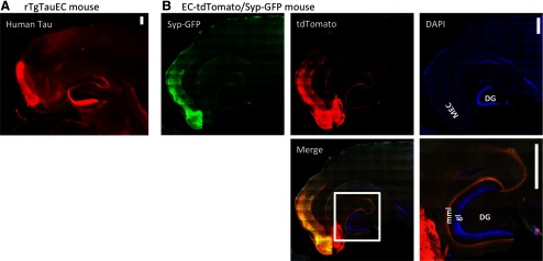
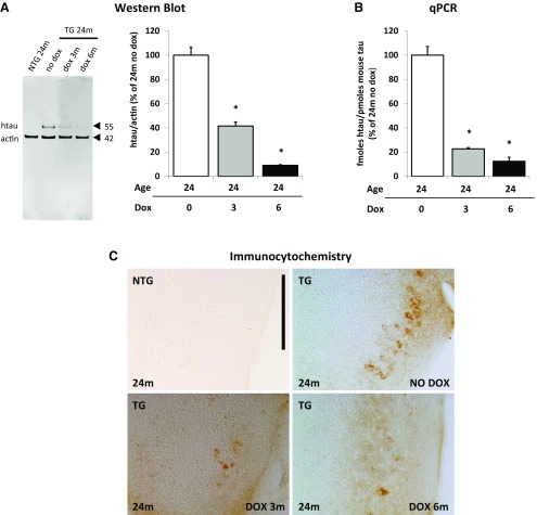
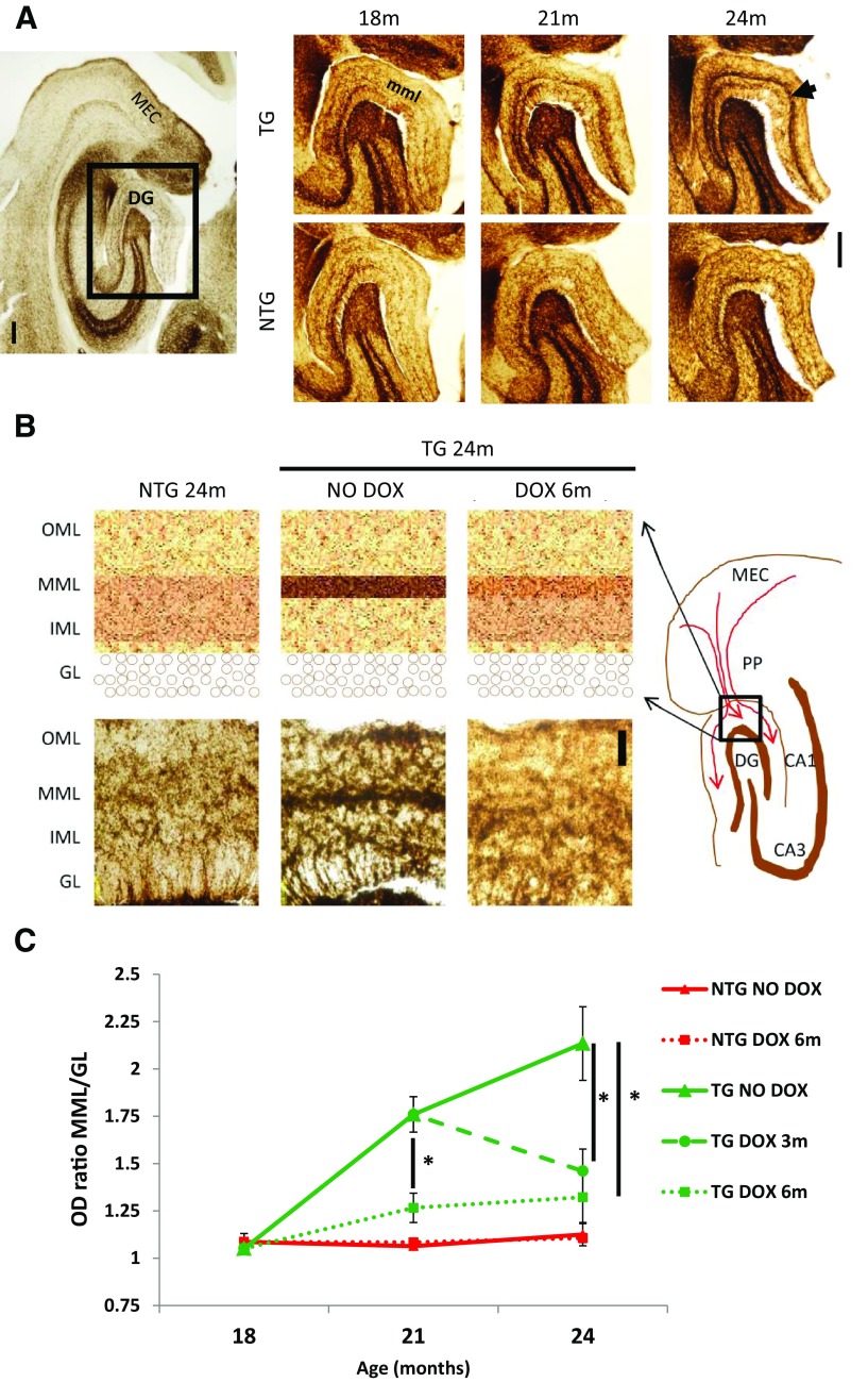
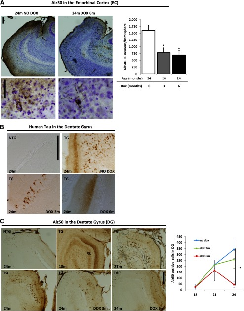
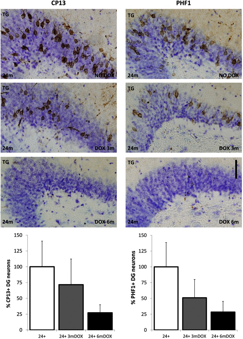
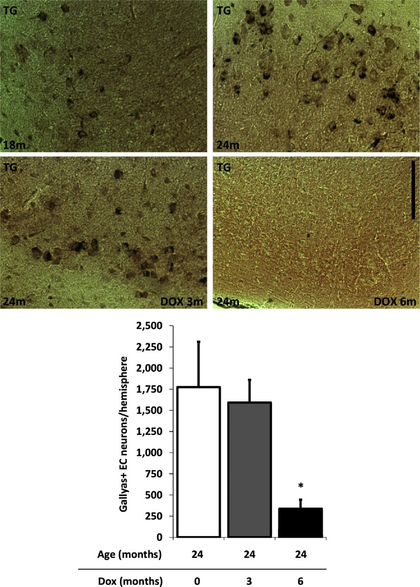
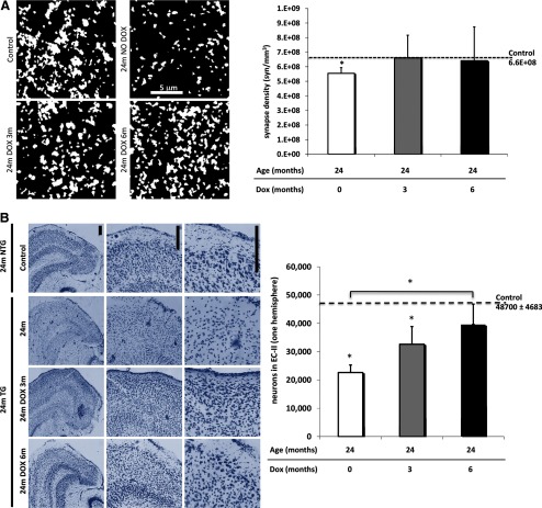
Neurofibrillary tangles (NFTs), a marker of neuronal alterations in Alzheimer's disease (AD) and other tauopathies, are comprised of aggregates of hyperphosphorylated tau protein. We recently studied the formation of NFTs in the entorhinal cortex (EC) and their subsequent propagation through neural circuits in the rTgTauEC mouse model (de Calignon et al., 2012). We now examine the consequences of suppressing transgene expression with doxycycline on the NFT-associated pathological features of neuronal system deafferentation, NFT progression and propagation, and neuronal loss. At 21 months of age we observe that EC axonal lesions are associated with an abnormal sprouting response of acetylcholinesterase (AChE)-positive fibers, a phenotype reminiscent of human AD. At 24 months, NFTs progress, tau inclusions propagate to the dentate gyrus, and neuronal loss is evident. Suppression of the transgene expression from 18 to 24 months led to reversal of AChE sprouting, resolution of Gallyas-positive and Alz50-positive NFTs, and abrogation of progressive neuronal loss. These data suggest that propagation of NFTs, as well as some of the neural system consequences of NFTs, can be reversed in an animal model of NFT-associated toxicity, providing proof in principle that these lesions can be halted, even in established disease.


Figures

Figure 1.
Figure 1.
Expression of P301L Tau restricted to the entorhinal cortex. rTgTauEC mice express human tau in neurons of the MEC that project to the middle molecular layer (mml) of the DG via the perforant pathway. These neurons are stained with a human tau antibody in A. To visualize the population of neurons expressing tau and where their synapses are located, we generated EC-tdTomato/Syp-GFP mice (B), which use the same neuropsin activator transgene to restrict expression to the EC crossed with a responder that expresses both myc-tagged tdTomato (red) in the entire cell and full-length mouse synaptophysin/mut4EGFP fusion protein (Syp-GFP) that localizes to synaptic vesicles (green); nuclei are labeled with DAPI (blue). Insert shows a higher-magnification image of the merged image. DG granule cells (gl) are marked by DAPI, Syn-GFP staining shows a fine discrete band in the middle molecular layer (mml) that colocalizes with the tdTomato labeling of the terminal zone of fibers from the EC cells (yellow). These mice confirm the expression of transgenes under this promoter is largely restricted to the EC and presubiculum and parasubiculum and that these neurons project axons that synapse in the middle molecular layer of the dentate gyrus. Importantly, they also confirm the absence of transgene expression in the DG granular cells Scale bars, 200 μm.
Figure 2.
Figure 2.
Tau expression in the medial entorhinal cortex is suppressed with doxycycline treatment. A–C, Doxycycline treatment suppressed transgene expression in animals at 24 months of age treated with doxycycline from 21 months of age for 3 months or from 18 months of age for 6 months. Results are labeled nontransgenic (NTG) and transgenic (TG). A, Western blot analysis using a human tau-specific antibody HT7 shows decreased levels of human tau protein (hTau) after 3 months and 6 months of DOX treatment. Levels of human tau protein were calculated by normalizing it to actin and are expressed as percentage of levels of hTau in 24-month-old animals that did not receive doxycycline treatment (n = 3 per group). *p = 0.005 and p = 0.008, respectively. B, The levels of human tau RNA were quantified by qPCR and showed reduced levels of RNA. The levels of hTau RNA were calculated by normalizing it to levels of endogenous mouse tau (n = 3 per group) and are expressed as percentage of levels of hTau in 24-month-old animals that did not receive doxycycline treatment. *p = 0.000147 and p = 0.00000057, respectively. C, Immunocytochemistry using HT7 confirmed a marked decrease in protein levels in the medial entorhinal cortex following transgene suppression. Scale bar, 200 μm. Results are expressed as the mean ± SEM.
Figure 3.
Figure 3.
Reorganization of AChE fibers in the perforant pathway terminal zone. Histochemistry for AChE-positive fibers in the middle molecular layer (mml) of dentate gyrus (DG) at 18, 21, and 24 months, in rTgTauEC mice and age-matched controls shows a deafferentation-induced AChE sprouting response at 21 and 24 months (A, arrow). Images are labeled nontransgenic (NTG) and transgenic (TG). Scale bar, 200 μm. A, Acetylcholinesterase staining showed sprouting of axon terminals from the inner molecular layer (IML) to a thinner and denser line in the middle molecular layer (MML). B shows a diagram representation of MEC, perforant pathway (PP), hippocampal regions CA1, CA3, and dentate gyrus (DG) with the region demarcated by black box (molecular layer) enlarged on the top left. Representative images of the molecular layer of the DG are shown on the bottom left. Scale bar, 50 μm. C, Comparing the ratio of optical densities between MML and IML reveals that the sprouting of AChE fibers is significant at 21 months of age and increases at 24 months, and that suppression of the transgene with a 3-month DOX treatment (n = 7) stops this synaptic reorganization when started at earlier age (p = 0.009) or at later age (p = 0.01). A 6-month treatment (n = 6) was more efficient (p = 0.006). Results are labeled nontransgenic (NTG) and transgenic (TG). Results are expressed as the mean ± SEM, *p < 0.05.
Figure 4.
Figure 4.
Transgene suppression reduces tau aggregation and propagation down neural circuits. A, Stereological counting of Alz50-positive neurons in the EC-II after transgene suppression shows that 3 and 6 months of DOX treatment significantly reduced the detection of Alz50-postive tau in the EC. This indicates that aggregation of misfolded tau in the entorhinal cortex is also prevented by 3 (p = 0.01751) and 6 months (p = 0.00973) of tau suppression. B, Higher magnification of these sections stained with Alz50 and cresyl violet illustrate the dense, punctate staining of misfolded tau in 24-month-old transgenic animals without DOX treatment, which is reduced with tau suppression. C, Immunocytochemistry using HT7 human tau-specific antibody shows a marked decrease in propagation of human tau protein to the dentate gyrus granular cells in animals at 24 months of age treated with doxycycline from 21 months of age for 3 months or from 18 months of age for 6 months. Spreading of tau pathology was quantified by Alz50-positive neuron counts in the granular layer (gl) of dentate gyrus (DG). Different groups of mice were treated with DOX either from 18 months of age for 3 months (n = 4) or for 6 months (n = 5); another group was treated from 21 months of age for 3 months (n = 8). No change in the extent of misfolded tau in the granule cells was evident after 3 months of doxycycline treatment. Longer treatment (6 months), however, significantly reduced the detection of human tau in the DG, showing that propagation of tau pathology to the dentate gyrus is reversed by 6 months of tau suppression (p = 0.005154). Images are labeled nontransgenic (NTG) and transgenic (TG). Scale bar, 200 μm. Results are expressed as the mean ± SEM, *p < 0.05.
Figure 5.
Figure 5.
Propagation of hyperphosphorylated tau to the dentate gyrus after 3 and 6 months of tau suppression. Propagation of hyperphosphorylated tau to the DG was assessed by CP13 (antibody against pSer202) and PHF1 (antibody against pSer396/404; PHF tau). There was no significant difference in the number of neurons labeled with CP13 by 3 months (n = 5; p = 0.26) and a slight trend toward reduction by 6 months (n = 5; p = 0.10) of tau suppression compared with 24-month untreated mice (n = 5). Similar results were observed for PHF1 with no significant changes by 3 months (n = 5; p = 0.14) and a slight trend toward reduction by 6 months (n = 5; p = 0.07) of tau suppression compared with 24-month untreated mice (n = 5). Sections were also stained with crystal violet. Images are labeled nontransgenic (NTG) and transgenic (TG). Results are expressed as the mean ± SEM, *p < 0.05. Scale bar, 50 μm.
Figure 6.
Figure 6.
Tangles, visualized by Gallyas silver staining, are reversed by 6 months of tau suppression. Most rTgTauEC animals develop silver-positive neurofibrillary tangles in the MEC by 18–21 months of age. Transgene suppression from 21 to 24 months (n = 5) did not prevent Gallyas-positive neurons from accumulating; however, treatment for 6 months from 18 to 24 months (n = 5) both prevented new tangles and reversed existing tangles as there were no Gallyas-positive cells in any 24-month-old animals treated for 6 months with DOX (p = 0.0386). Images are labeled nontransgenic (NTG) and transgenic (TG). Scale bar, 100 μm. Results are expressed as the mean ± SEM, *p < 0.05.
Figure 7.
Figure 7.
Synaptic loss in the target zone of the perforant pathway and neuronal loss in MEC can be prevented by doxycycline treatment. A, Array tomography using synaptophysin to label presynaptic structures in the middle molecular layer of the DG (scale bar, 5 μm) shows presynaptic loss at 24 months of age, indicating loss of synapses between EC-II neurons and DG neurons, which is prevented by 6 months of tau suppression (p = 0.0233). Images are maximum intensity projections of 15 × 15 μm regions of interest from 10 serial sections (70 nm thickness) from processed output files showing objects counted as synapses. B, Neuronal counts were performed on transgenic and control animals at 24 months of age treated with doxycycline starting either from 21 months of age for 3 months or from 18 months of age for 6 months, using stereological estimations of cresyl violet-labeled neuronal nuclei (n = 5 per group). rTgTauEC mice showed decreased neuronal counts at 24 months of age compared with controls (p < 0.0001), which is ameliorated by 3 months of tau suppression (p = 0.0499) and prevented by 6 months of transgene suppression (p = 0.0009). Representative images of cresyl violet labeled nuclei of the entorhinal cortex (EC) are shown on top; scale bar, 200 μm. There was partial recovery of neuronal loss in the EC after 6 months of doxycycline treatment. Results are expressed as the mean ± SEM, *p < 0.05.

References

    1. Aubert I, Poirier J, Gauthier S, Quirion R. Multiple cholinergic markers are unexpectedly not altered in the rat dentate gyrus following entorhinal cortex lesions. J Neurosci. 1994;14:2476–2484. - PMC - PubMed
    1. Berger Z, Roder H, Hanna A, Carlson A, Rangachari V, Yue M, Wszolek Z, Ashe K, Knight J, Dickson D, Andorfer C, Rosenberry TL, Lewis J, Hutton M, Janus C. Accumulation of pathological tau species and memory loss in a conditional model of tauopathy. J Neurosci. 2007;27:3650–3662. doi: 10.1523/JNEUROSCI.0587-07.2007. - DOI - PMC - PubMed
    1. Braak H, Braak E. Neuropathological stageing of Alzheimer-related changes. Acta Neuropathol. 1991;82:239–259. doi: 10.1007/BF00308809. - DOI - PubMed
    1. Braak H, Thal DR, Ghebremedhin E, Del Tredici K. Stages of the pathologic process in Alzheimer disease: age categories from 1 to 100 years. J Neuropathol Exp Neurol. 2011;70:960–969. doi: 10.1097/NEN.0b013e318232a379. - DOI - PubMed
    1. Carrettiero DC, Hernandez I, Neveu P, Papagiannakopoulos T, Kosik KS. The cochaperone BAG2 sweeps paired helical filament- insoluble tau from the microtubule. J Neurosci. 2009;29:2151–2161. doi: 10.1523/JNEUROSCI.4660-08.2009. - DOI - PMC - PubMed

Publication types