. 2013 Aug 14;33(33):13410-24.
doi: 10.1523/JNEUROSCI.1534-13.2013.

Rescue of peripheral and CNS axon defects in mice lacking NMNAT2

Affiliations

Rescue of peripheral and CNS axon defects in mice lacking NMNAT2

Jonathan Gilley et al. J Neurosci. .

Abstract

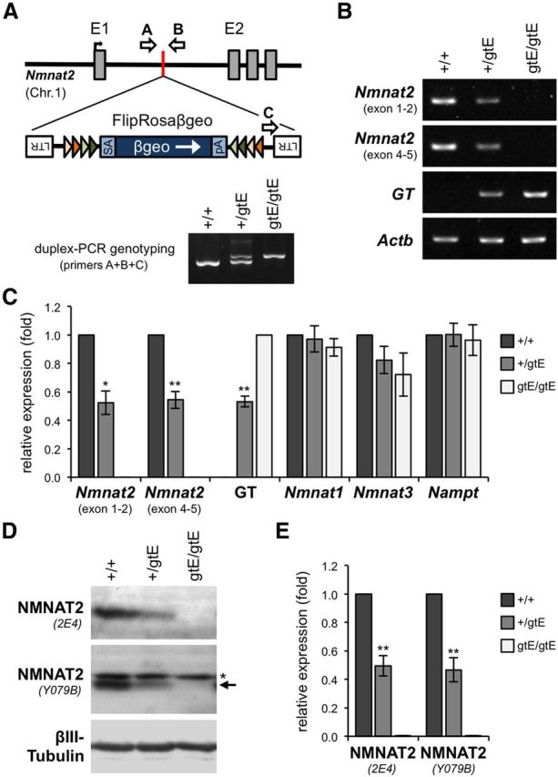
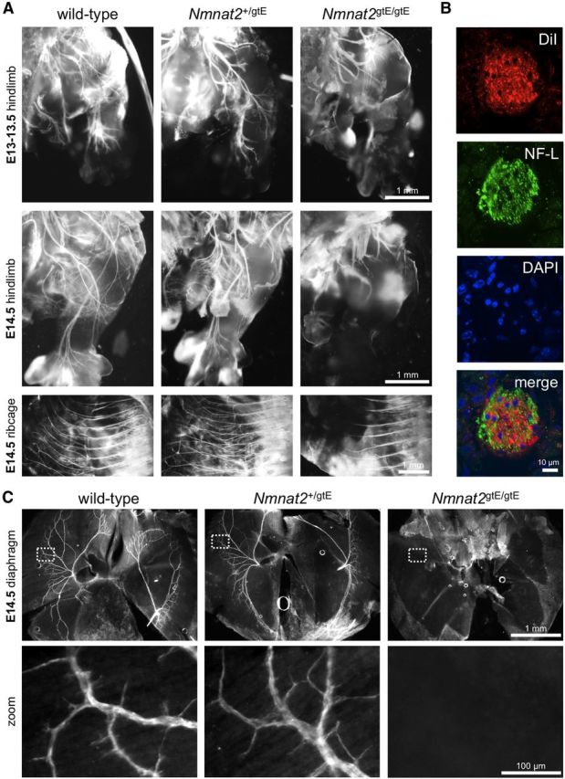
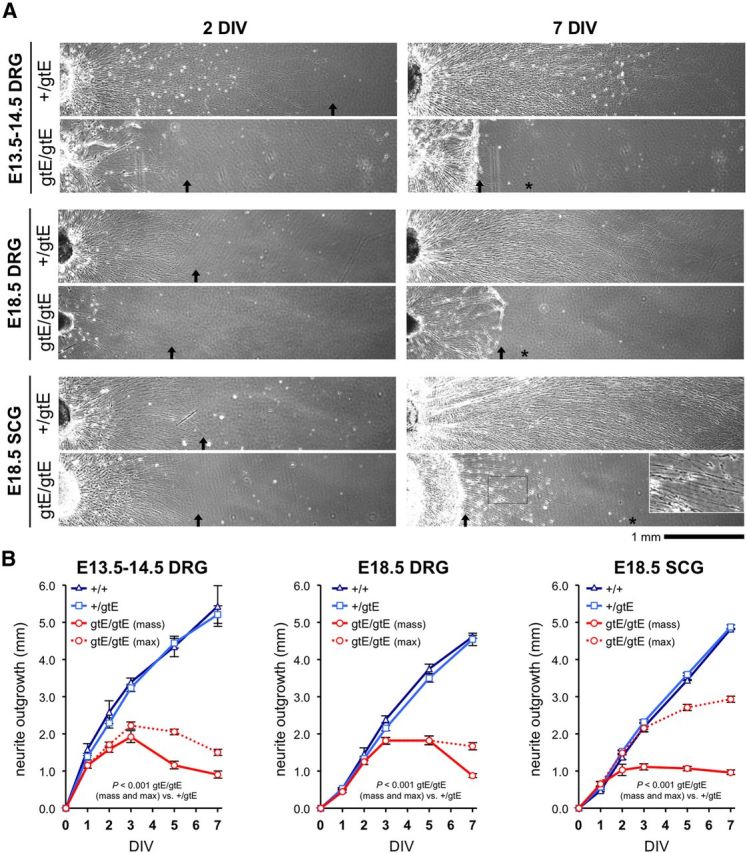
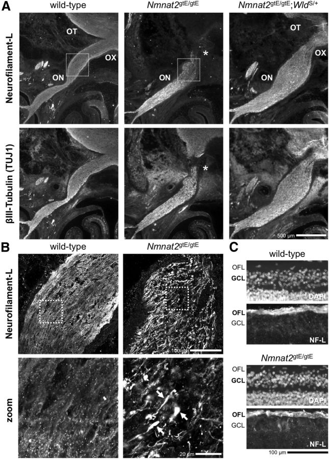
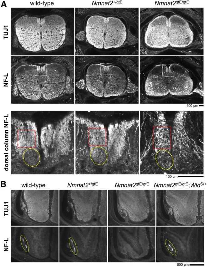
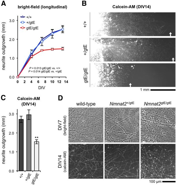
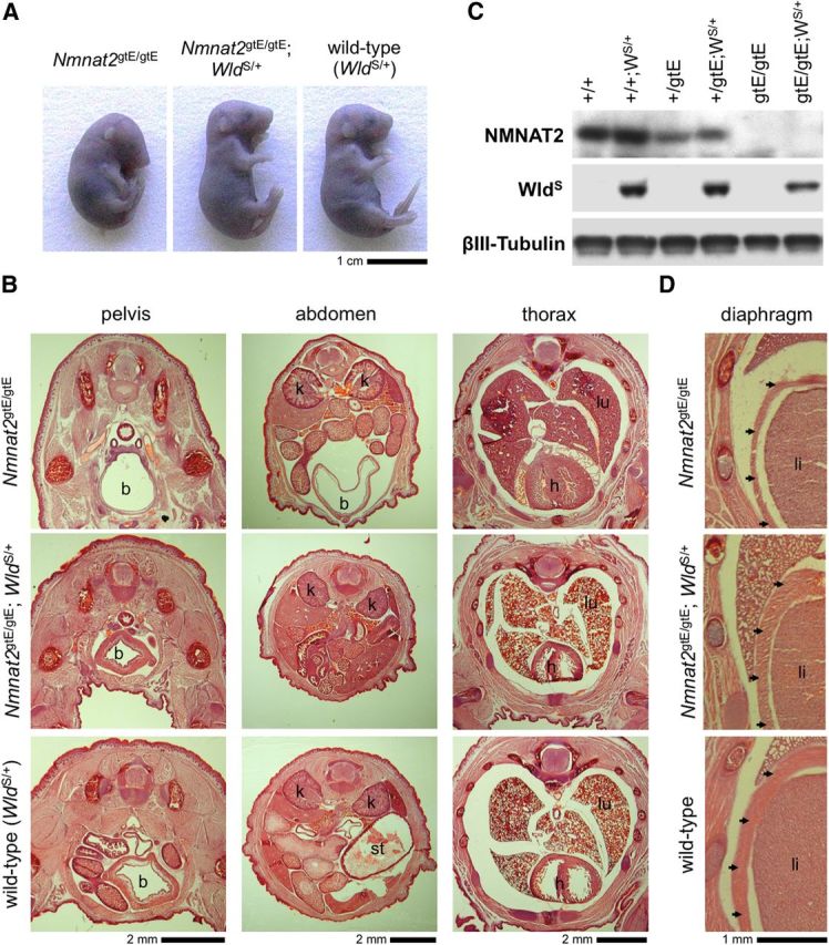
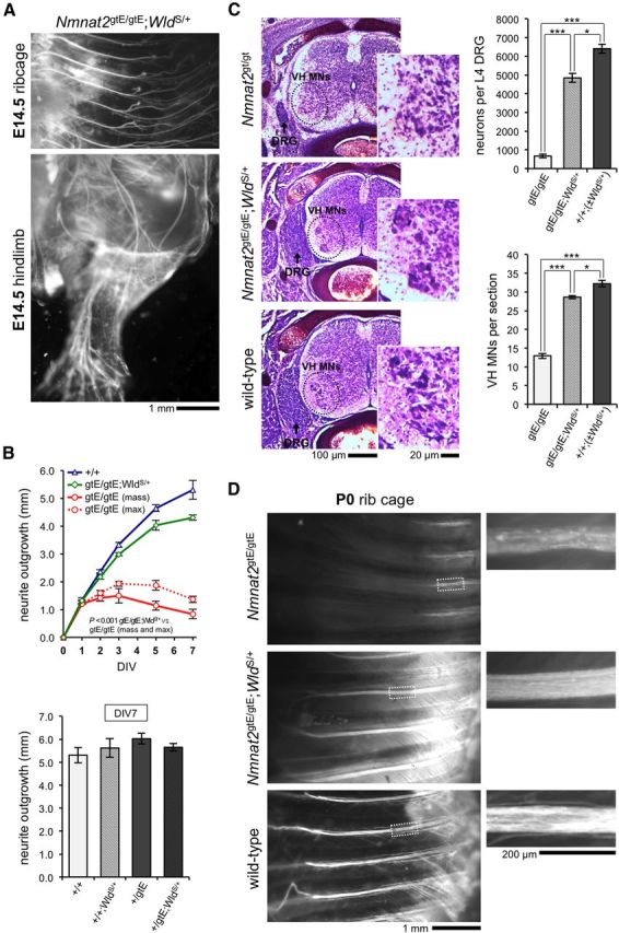
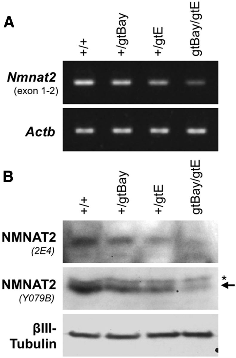
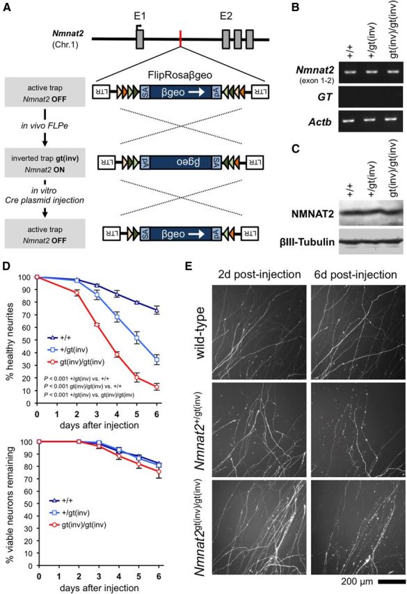
NMNAT2 is an NAD(+)-synthesizing enzyme with an essential axon maintenance role in primary culture neurons. We have generated an Nmnat2 gene trap mouse to examine the role of NMNAT2 in vivo. Homozygotes die perinatally with a severe peripheral nerve/axon defect and truncated axons in the optic nerve and other CNS regions. The cause appears to be limited axon extension, rather than dying-back degeneration of existing axons, which was previously proposed for the NMNAT2-deficient Blad mutant mouse. Neurite outgrowth in both PNS and CNS neuronal cultures consistently stalls at 1-2 mm, similar to the length of truncated axons in the embryos. Crucially, this suggests an essential role for NMNAT2 during axon growth. In addition, we show that the Wallerian degeneration slow protein (Wld(S)), a more stable, aberrant NMNAT that can substitute the axon maintenance function of NMNAT2 in primary cultures, can also correct developmental defects associated with NMNAT2 deficiency. This is dose-dependent, with extension of life span to at least 3 months by homozygous levels of Wld(S) the most obvious manifestation. Finally, we propose that endogenous mechanisms also compensate for otherwise limiting levels of NMNAT2. This could explain our finding that conditional silencing of a single Nmnat2 allele triggers substantial degeneration of established neurites, whereas similar, or greater, reduction of NMNAT2 in constitutively depleted neurons is compatible with normal axon growth and survival. A requirement for NMNAT2 for both axon growth and maintenance suggests that reduced levels could impair axon regeneration as well as axon survival in aging and disease.


Figures

Figure 1.
Figure 1.
Generation and initial characterization of NMNAT2-deficient mice. A, Localization of the conditional-ready gene trap within intron 1 of the mouse Nmnat2 gene in mice derived from EUCOMM ES cell clone EUCE0262a08. Features are as described previously (Schnutgen et al., 2005). Positions of PCR genotyping primers (arrows A–C), and representative genotyping results for wild-type (+/+), Nmnat2+/gtE, and Nmnat2gtE/gtE animals, are also shown. B, Representative RT-PCR showing Nmnat2 mRNA levels (two sets of primers), levels of the mRNA derived from trapping (GT), and Actb mRNA levels (acting as a reference), in brains of wild-type (+/+), Nmnat2+/gtE, and Nmnat2gtE/gtE E18.5 embryos. C, Quantification of RT-PCR data showing relative mean levels (± SEM) of Nmnat2, GT, Nmnat1, Nmnat3, and Nampt mRNAs after normalization to Actb mRNA (n = 4 for each genotype). Data are presented relative to wild-type levels (set at 1), except for GT data, which is presented relative to Nmnat2gtE/gtE levels. *p < 0.05 (two-tailed one sample t test). **p < 0.01 (two-tailed one sample t test). D, Representative immunoblots showing Nmnat2 levels (two antibodies) in brains of wild-type (+/+), Nmnat2+/gtE, and Nmnat2gtE/gtE E18.5 embryos. βIII-Tubulin acts as the sample control. NMNAT2 migrates at ∼32 kDa. A nonspecific band (*), slightly larger than NMNAT2 (arrow), is detected by Y079B. E, Quantification of immunoblot data showing relative mean levels (± SEM) of NMNAT2 detected with each antibody after normalization to βIII-Tubulin (n = 4 for each genotype). Data are presented relative to wild-type levels (set at 1). **p < 0.01 (two-tailed one sample t test).
Figure 2.
Figure 2.
DiI labeling and neurofilament immunostaining reveal peripheral nerve defects in E13–E14.5 Nmnat2gtE/gtE embryos. A, DiI-labeled spinal nerve branches in the hindlimb and intercostal nerves in the ribcages of E13/13.5 and E14.5 wild-type, Nmnat2+/gtE, and Nmnat2gtE/gtE embryos 5 weeks after insertion of DiI crystals into the thoracic and subthoracic spinal cord (images are representative of at least n = 3 embryos of each genotype). B, Confocal optical slice from a transverse cryosection of an intercostal nerve immunostained for NF-L after diffusion of DiI from the thoracic spinal cord of a wild-type E18.5 embryo confirms labeling of structures within the nerve by DiI (DAPI counterstaining reveals nuclei). Lack of precise colocalization is consistent with DiI labeling membranes and NF-L labeling the cytoskeleton. C, Whole-mount NF-L immunostaining of diaphragms from wild-type, Nmnat2+/gtE, and Nmnat2gtE/gtE E14.5 embryos (representative of n = 3 for each genotype) to label phrenic nerve branches. No branches were detected in Nmnat2gtE/gtE diaphragms. NF-H immunostaining revealed a similar lack of phrenic nerve branches in Nmnat2gtE/gtE E14.5 embryos (n = 2 for each genotype), although staining was weaker because of low levels of NF-H expression at this stage (data not shown).
Figure 3.
Figure 3.
Restricted neurite outgrowth in explant cultures of Nmnat2gtE/gtE DRGs and SCGs at different embryonic stages. A, Representative images of neurite outgrowth at 2 DIV and 7 DIV from cultured E13.5–E14.5 and E18.5 DRG explants and E18.5 SCG explants from Nmnat2+/gtE and Nmnat2gtE/gtE embryos. Nmnat2+/gtE explants were indistinguishable from wild-type explants (data not shown) in all cases (see B). Nmnat2gtE/gtE DRGs taken from E18.5 embryos were noticeably smaller than wild-type and Nmnat2+/gtE DRGs, reflecting extensive DRG loss that occurs at later developmental stages in Nmnat2gtE/gtE embryos (Fig. 8C). The limit of mass outgrowth of the majority of neurites is indicated by an arrow (if located in the image shown) and maximum extension of smaller numbers of neurites in Nmnat2gtE/gtE cultures by an asterisk. Higher magnification of the boxed region in the Nmnat2gtE/gtE SCG culture at 7 DIV shows deteriorating health of the small population of longer neurites. B, Mean radial neurite outgrowth (mm ± SEM) up to 7 DIV in DRG and SCG explant cultures (as indicated) from wild-type (+/+), Nmnat2+/gtE, and Nmnat2gtE/gtE E14.5 or E18.5 embryos. Mass outgrowth (mean extension of the majority of neurites) and maximum (max) outgrowth (mean extension of smaller populations of longer neurites) are plotted separately for Nmnat2gtE/gtE cultures. Each analysis involved n = 2 wild-type, n = 7–11 Nmnat2+/gtE, and n = 6 Nmnat2gtE/gtE embryos, with average growth determined from 1 to 3 ganglia for each embryo. p values were calculated from embryo averages using two-way repeated-measures ANOVA with Bonferroni post hoc correction. Wild-type and Nmnat2+/gtE growth curves were essentially indistinguishable in all cases.
Figure 4.
Figure 4.
An RGC axon defect in Nmnat2gtE/gtE optic nerve. A, Low-magnification wide-field images of NF-L and βIII-Tubulin (TUJ1) immunostaining of horizontal cryosections of the heads of wild-type, Nmnat2gtE/gtE, and Nmnat2gtE/gtE;WldS/+ E18.5 embryos at the level of the optic nerve (ON), optic chiasma (OX), and optic tract (OT). Maximal length of NF-L and βIII-Tubulin-labeled RGC axons in the Nmnat2gtE/gtE ON (as revealed by staining of adjacent serial sections for >150 μm in each direction) is indicated by an asterisk. The RGC axon defect appears to be largely corrected in Nmnat2gtE/gtE;WldS/+ embryos. B, Confocal slices of the boxed regions in A highlight abnormal swellings containing accumulations of NF-L at the distal ends of the truncated RGC axons in Nmnat2gtE/gtE embryos. Arrows point to swellings in higher-magnification images (zoom) of the boxed regions in the panels above. C, NF-L immunostaining and DAPI counterstaining of horizontal sections through the retinas of wild-type and Nmnat2gtE/gtE E18.5 embryos revealed a normal optic fiber layer (OFL) and no evidence of significant pyknosis in the ganglion cell layer (GCL) in the mutant. Images are representative of n = 3 embryos of each genotype. Nmnat2+/gtE embryos (n = 3) were indistinguishable from wild-types (data not shown).
Figure 5.
Figure 5.
Axon defects in the spinal cord and olfactory bulb in E18.5 Nmnat2gtE/gtE embryos. Images of NF-L and βIII-Tubulin (TUJ1) immunostaining of horizontal cryosections of the lower cervical spinal cord (and surrounding tissues) (A), and the olfactory bulb (B), of wild-type, Nmnat2+/gtE, Nmnat2gtE/gtE, and Nmnat2gtE/gtE;WldS/+ E18.5 embryos (representative of serial sections from n = 3 embryos of each genotype). Higher-magnification images of NF-L immunostaining in the dorsal column of the spinal cord (white boxed region) are shown at the bottom of A. The approximate locations of the corticospinal tract (yellow circle) and gracile tract (red box) are indicated. The dorsal column appears elongated in the Nmnat2gtE/gtE embryo, but the overall area is similar to that in wild-type and Nmnat2+/gtE embryos. However, there is a clear deficiency of axons in the corticospinal tract in the Nmnat2gtE/gtE dorsal column, and the gracile tract appears to contain fewer, severely dystrophic axons. B, The olfactory nerve layer (circled in yellow) can easily be identified in wild-type, Nmnat2+/gtE, and Nmnat2gtE/gtE;WldS/+ sections but is absent from serial sections through the comparable region in Nmnat2gtE/gtE embryos.
Figure 6.
Figure 6.
Restricted neurite outgrowth in Nmnat2gtE/gtE cortical explant cultures. A, Mean radial neurite outgrowth (mm ± SEM) up to 14 DIV in cortical explant cultures from wild-type (+/+), Nmnat2+/gtE, and Nmnat2gtE/gtE E18.5 embryos (n = 4 embryos per genotype, with average growth per embryo determined from 2 separate explants). p values were calculated from embryo averages using two-way repeated-measures ANOVA with Bonferroni post hoc correction. B, Representative fluorescent images of cortical explant cultures stained with calcein-AM at 14 DIV. C, Mean radial neurite outgrowth (mm ± SEM) in cortical explant cultures as revealed by Calcein-AM staining at 14 DIV. Data are from the same cultures as in A. **p < 0.01, Nmnat2gtE/gtE versus both wild-type and Nmnat2+/gtE (calculated from embryo averages using one-way ANOVA with Bonferroni post hoc correction). Measurements based on calcein-AM staining and phase-contrast images are therefore consistent with each other. D, Representative phase-contrast images of neurites in cortical explant cultures at 7 DIV, and fluorescent images of calcein-AM stained neurites at 14 DIV. Significant neurite blebbing is visible in Nmnat2gtE/gtE cultures at both time points.
Figure 7.
Figure 7.
Gross morphological defects found in newborn pups lacking NMNAT2 are rescued by WldS expression. A, Posture of Nmnat2gtE/gtE, Nmnat2gtE/gtE;WldS/+, and wild-type (WldS/+) E18.5 embryos. All Nmnat2gtE/gtE;WldS/+ E18.5 embryos and newborn pups observed to date (n = 13) had a normal posture. Heterozygosity for WldS does not affect the gross appearance of wild-type or Nmnat2+/gtE embryos (which are also normal; data not shown). B, Hematoxylin and eosin-stained pelvic, abdominal, and thoracic cryosections from newborn Nmnat2gtE/gtE, Nmnat2gtE/gtE;WldS/+, and wild-type (WldS/+) pups. A distended bladder (b) can be seen to extend to the level of the kidneys (k) in the abdomen of the Nmnat2gtE/gtE pup. There is also reduced muscle mass in the thigh, pelvis, and abdominal walls, and collapsed lungs (lu) are indicative of a failure to breathe. The Nmnat2gtE/gtE;WldS/+ pup appears similar to the wild-type with a nondistended bladder, normal muscle mass, and lungs (lu) that show clear signs of inflation. A distended stomach (st) can be seen in the abdomen of the wild-type pup. C, Immunoblot showing selective expression of WldS in newborn wild-type (+/+), Nmnat2+/gtE, and Nmnat2gtE/gtE pups heterozygous for WldS (WS/+). βIII-Tubulin represents the sample control, and NMNAT2 levels confirm the Nmnat2 genotype. D, Hematoxylin and eosin-stained cryosections showing an underdeveloped diaphragm in a newborn Nmnat2gtE/gtE pup but normal diaphragms in newborn Nmnat2gtE/gtE;WldS/+ and wild-type pups. The diaphragm (arrowhead) lies between the liver (li), the lungs (top), and the ribcage (left) in these sections. B, D, Images are representative of at least n = 3 per genotype.
Figure 8.
Figure 8.
Whole nerve and neuronal defects found in newborn pups lacking NMNAT2 are rescued by WldS expression. A, DiI-labeled intercostal nerves in the ribcage and spinal nerve branches in the hindlimb of an Nmnat2gtE/gtE;WldS/+ E14.5 embryo (representative of n = 3 and matching images in Fig. 2A). B, Mean radial neurite outgrowth (mm ± SEM) up to 7 DIV in E14.5 DRG explant cultures from wild-type (+/+), Nmnat2gtE/gtE, and Nmnat2gtE/gtE;WldS/+ embryos (top: n = 2, 5, and 3 embryos, respectively, with average growth per embryo determined from 1–3 DRGs), and at 7 DIV only in cultures from Nmnat2+/gtE embryos ± WldS/+ (bottom: n = 2 or 3 embryos per genotype/3 DRGs per embryo). p values (top) were calculated from embryo averages using two-way repeated-measures ANOVA with Bonferroni post hoc correction. Data are independent of that in Figure 3B. WldS/+ does not significantly affect wild-type and Nmnat2+/gtE DRG neurite outgrowth (bottom: one-way ANOVA). C, Nissl-stained horizontal cryosections at the level of the L3 DRG (widest aspect), from newborn Nmnat2gtE/gtE, Nmnat2gtE/gtE;WldS/+, and wild-type;WldS/+ (+/+;WldS/+) pups. Reduced DRG size (arrowed) and reduced numbers of ventral horn motor neurons (VH MNs) in the spinal cord (magnified for clarity) are evident in the Nmnat2gtE/gtE pup and are rescued in the Nmnat2gtE/gtE;WldS/+ pup. Mean neuron numbers (± SEM) in L4 DRGs (top) and mean numbers (± SEM) of VH MNs per section in lower lumbar spinal cord (bottom) in each genotype. Data are from n = 3 or 4 pups for each genotype (wild-types ± WldS/+ were grouped; n = 2 each). ***p < 0.001 (one-way ANOVA with Bonferroni post hoc correction). *p < 0.05 (one-way ANOVA with Bonferroni post hoc correction). D, DiI-labeled intercostal nerves in the ribcages of newborn Nmnat2gtE/gtE, Nmnat2gtE/gtE;WldS/+, and wild-type pups (representative of n = 3 per genotype). Magnified regions highlight axon degeneration in Nmnat2gtE/gtE intercostal nerves.
Figure 9.
Figure 9.
Subheterozygous levels of NMNAT2 expression in Nmnat2gtBay/gtE compound heterozygote mice. A, RT-PCR showing levels of Nmnat2 mRNA in brains of 6 month wild-type, Nmnat2+/gtBay, Nmnat2+/gtE, and Nmnat2gtBay/gtE mice. Levels of Actb mRNA act as a reference. B, Representative immunoblot showing NMNAT2 levels (two independent antibodies) in brains of 6 month wild-type, Nmnat2+/gtBay, Nmnat2+/gtE, and Nmnat2gtBay/gtE mice. βIII-Tubulin acts as the sample control.
Figure 10.
Figure 10.
Silencing of a single Nmnat2 allele is sufficient to induce neurite degeneration in primary SCG neuron cultures. A, Schematic showing how sequential exposure to FLPe and Cre recombinases allows restoration of expression from the targeted Nmnat2 allele, and subsequent resilencing, through successive inversion of the gene trap facilitated by pairs of inversely orientated heterotypic FLPe and Cre recombinase target sequences (colored triangles) (Schnutgen et al., 2005). B, C, Representative RT-PCR (B) and representative immunoblot (C) showing Nmnat2 mRNA and NMNAT2 protein levels in brains of wild-type, Nmnat2+/gt(inv), and Nmnat2gt(inv)/gt(inv) P1 pups. Actb and βIII-Tubulin represent sample references. D, Survival of DsRed2-labeled distal neurites (top) and neuron viability (bottom) for wild-type (+/+), Nmnat2+/gt(inv), and Nmnat2gt(inv)/gt(inv) SCG neurons injected with a Cre-EGFP expression vector and pDsRed2 (both at 10 ng/μl). The mean percentage (± SEM) of healthy DsRed2-labeled neurites or viable cell bodies remaining each day after injection, of those (both healthy and abnormal) present in the same field at 2 d after injection, is plotted. Neurite data are from multiple fields in five independent injection experiments for each genotype. Injected cell bodies with normal gross morphology were classed as viable. We previously demonstrated that morphology revealed by fluorescent proteins is a reliable indicator of neuronal viability (Gilley and Coleman, 2010). p values were calculated from individual experiment averages using two-way repeated-measures ANOVA with Bonferroni post hoc correction. We also saw no increase in neurite degeneration (relative to wild-type) for Nmnat2+/gt(inv) and Nmnat2gt(inv)/gt(inv) SCG neurons injected with a control EGFP expression vector, rather than Cre-EGFP (data not shown). E, Representative fields of DsRed2-labeled distal neurites from experiments quantified in D at 2 and 6 d after injection.

Comment in

References

    1. Adalbert R, Coleman MP. Axon pathology in age-related neurodegenerative disorders. Neuropathol Appl Neurobiol. 2012 doi: 10.1111/j.1365-2990.2012.01308.x. doi: 10.1111/j.1365-2990.2012.01308.x. Advance online publication. Retrieved Oct. 10, 2012. - DOI - PubMed
    1. Babetto E, Beirowski B, Russler EV, Milbrandt J, Diantonio A. The Phr1 ubiquitin ligase promotes injury-induced axon self-destruction. Cell Rep. 2013;3:1422–1429. doi: 10.1016/j.celrep.2013.04.013. - DOI - PMC - PubMed
    1. Berger F, Lau C, Dahlmann M, Ziegler M. Subcellular compartmentation and differential catalytic properties of the three human nicotinamide mononucleotide adenylyltransferase isoforms. J Biol Chem. 2005;280:36334–36341. doi: 10.1074/jbc.M508660200. - DOI - PubMed
    1. Bhattacharya MR, Gerdts J, Naylor SA, Royse EX, Ebstein SY, Sasaki Y, Milbrandt J, DiAntonio A. A model of toxic neuropathy in Drosophila reveals a role for MORN4 in promoting axonal degeneration. J Neurosci. 2012;32:5054–5061. doi: 10.1523/JNEUROSCI.4951-11.2012. - DOI - PMC - PubMed
    1. Bowers CW, Zigmond RE. Localization of neurons in the rat superior cervical ganglion that project into different postganglionic trunks. J Comp Neurol. 1979;185:381–391. doi: 10.1002/cne.901850211. - DOI - PubMed

Publication types

MeSH terms

Substances