. 2013 Aug 14;33(33):13475-83.
doi: 10.1523/JNEUROSCI.0419-13.2013.

Therapeutic margins in a novel preclinical model of retinitis pigmentosa

Affiliations

Therapeutic margins in a novel preclinical model of retinitis pigmentosa

Richard J Davis et al. J Neurosci. .

Abstract

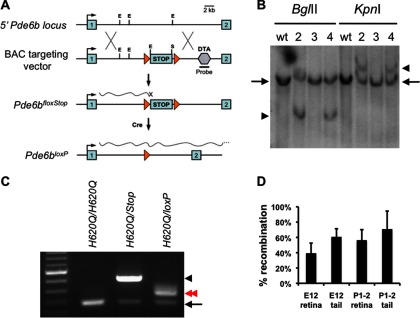
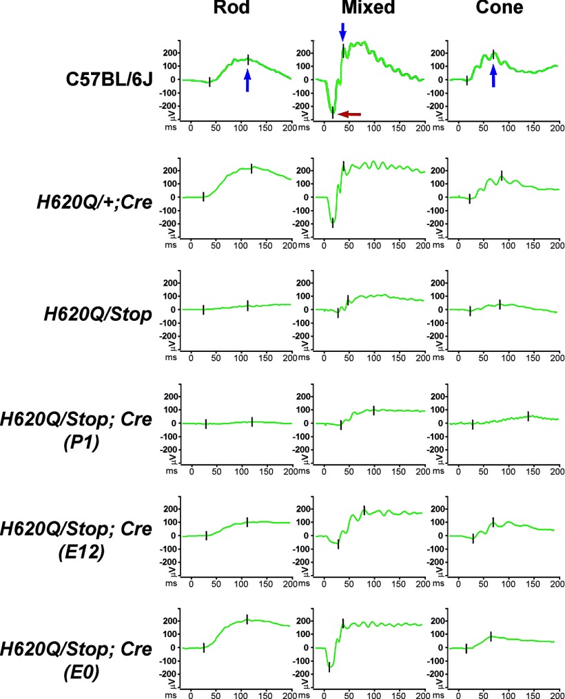
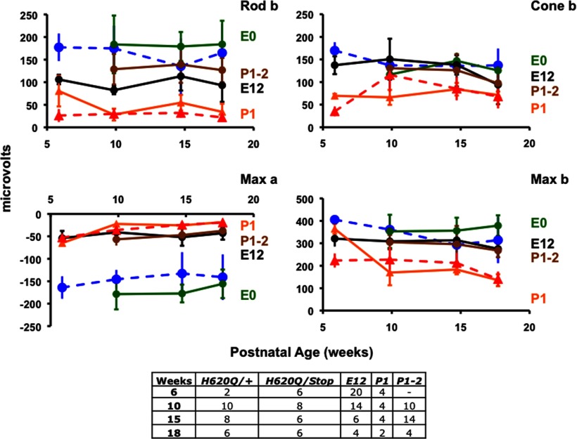
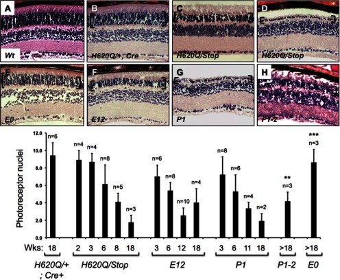
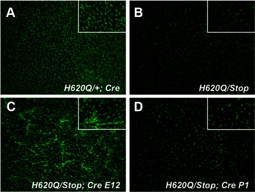
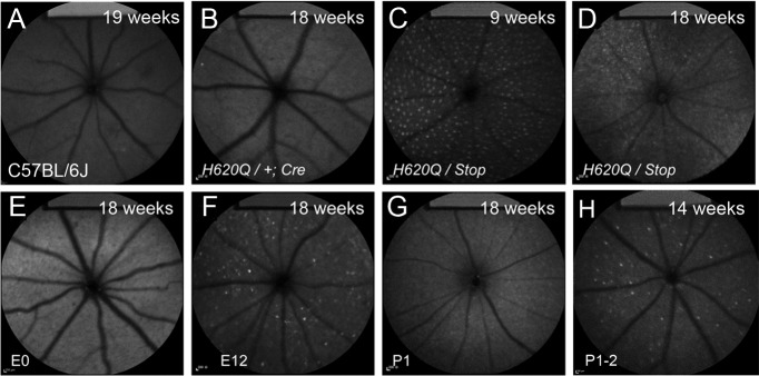
The third-most common cause of autosomal recessive retinitis pigmentosa (RP) is due to defective cGMP phosphodiesterase-6 (PDE6). Previous work using viral gene therapy on PDE6-mutant mouse models demonstrated photoreceptors can be rescued if administered before degeneration. However, whether visual function can be rescued after degeneration onset has not been addressed. This is a clinically important question, as newly diagnosed patients exhibit considerable loss of rods and cones in their peripheral retinas. We have generated and characterized a tamoxifen inducible Cre-loxP rescue allele, Pde6b(Stop), which allows us to temporally correct PDE6-deficiency. Whereas untreated mutants exhibit degeneration, activation of Cre-loxP recombination in early embryogenesis produced stable long-term rescue. Reversal at later time-points showed partial long-term or short-lived rescue. Our results suggest stable restoration of retinal function by gene therapy can be achieved if a sufficient number of rods are treated. Because patients are generally diagnosed after extensive loss of rods, the success of clinical trials may depend on identifying patients as early as possible to maximize the number of treatable rods.


Figures

Figure 1.
Figure 1.
Production of a reversible Pde6bStop allele, and Cre-mediated generation of Pde6bloxP. A, Scheme for generating the Cre-inducible Pde6bStop allele. Pde6b was targeted with a BAC vector (clone RP23 131C17) containing exons 1 and 2, a floxed Stop/Pgk-neo cassette (loxP triangles flanking STOP), and a DTA-negative selection cassette (hexagon). Homologous recombination places Stop in intron 1 thereby blocking Pde6b expression (wavy line with X). Cre-mediated recombination removes the Stop cassette restoring expression (wavy long line). Recombination was detected in ES cells using a probe sequence that is deleted by introduction of the DTA cassette and is absent in the targeting vector. B, Identification of the Pde6bStop allele. Southern analysis of ES cell lines 2 and 4 with an intron 1 probe (not present in targeting vector) detects wild-type (arrows) and recombinant allele (arrowheads) fragments. Line 3 is a nonrecombinant line. C, Cre-mediated recombination detection by PCR. A three-primer assay distinguishes Stop (arrowhead), and loxP (red arrow) alleles within intron 1 (see Materials and Methods). The presence of the H620Q mutation is confirmed by both wild-type sequences in intron 1 and DNA sequencing of exon 15 (black arrow). D, Detection of Stop allele recombination in H620Q/Stop; Rosa26CreERT2 tails and retinas after tamoxifen treatment at E12 or P1–P2 (see Materials and Methods). Percentage recombination is expressed as the amplicon ratio: loxP/(loxP+Stop) ×100%.
Figure 2.
Figure 2.
Examples of ERG traces from control and experimental Pde6bH620Q/Pde6bStop mice at 18 weeks. Scotopic rod-specific (rod) and mixed rod–cone (mixed), and photopic cone (cone) responses (green traces). Top panels, blue arrows indicate b-wave peaks, whereas the red arrow indicates an a-wave peak, which is also shown as tick marks. Average peak values were calculated for eye on each mouse as a function of age and combined into the graph shown in Figure 3.
Figure 3.
Figure 3.
Rescue of scotopic and photopic function after Stop-loxP recombination. Scotopic rod-specific (rod b) and mixed rod–cone (max a, max b), and photopic cone (cone b) responses in positive control Pde6bH620Q/Pde6b+ (dashed blue) and mutant Pde6bH620/Pde6bStop mice (dashed red). Ages of mice in postnatal weeks on x-axis. Experimental mice (H620Q/Stop; Rosa26CreERT2) E0 (green line), E12 (black line), P1 (orange line), and P1–P2 (brown line). Averages and SDs of photoreceptor (a-wave) or inner retina mediated (b-wave) peak intensities are plotted in microvolts on the y-axis. Number of eyes assayed is shown in Table 1.
Figure 4.
Figure 4.
Rescue of Pde6bH620Q/Pde6bStop photoreceptor cells rescue after Cre-loxP recombination. H&E stained retinal sections from wild-type (A), positive control (B), mutant control (C, D), and experimental E0, E12, or P1 mice (E–H). All panels are from 18-week-old mice, except D, which is from a 9-week-old mouse. The ONL is indicated with brackets. F, Quantitative analysis of the number of rows of photoreceptor nuclei in histological sections per week. Averages and SDs were calculated from multiple retinas (n is shown above each column). Unpaired two-tailed Student's t tests were performed on mutant control and experimental data. *p < 0.05, **p < 0.01, ***p < 0.001.
Figure 5.
Figure 5.
Partial rescue of Pde6bH620Q/Pde6bStop cone dysmorphology. PNA-stained flat mount retinas from 18-week-old positive control (A), mutant control (B), experimental E12 (C), and P1 (D) mice. PNA is conjugated to Alexa488. Insets show higher magnification.
Figure 6.
Figure 6.
Partial rescue of Pde6bH620Q/Pde6bStop fundus autofluorescence phenotype. Fundus images from C57BL/6J control (A), positive control (B), mutant control (C, D), and experimental E0, E12, P1, or P1–P2 mice (E–H). All panels are from 18-week-old mice, except C, which is from a 9-week-old mouse.

References

    1. Allocca M, Manfredi A, Iodice C, Di Vicino U, Auricchio A. AAV-mediated gene replacement, either alone or in combination with physical and pharmacological agents, results in partial and transient protection from photoreceptor degeneration associated with betaPDE deficiency. Invest Ophthalmol Vis Sci. 2011;52:5713–5719. doi: 10.1167/iovs.10-6269. - DOI - PubMed
    1. Badea TC, Wang Y, Nathans J. A noninvasive genetic/pharmacologic strategy for visualizing cell morphology and clonal relationships in the mouse. J Neurosci. 2003;23:2314–2322. - PMC - PubMed
    1. Berson EL. Retinitis pigmentosa: the Friedenwald lecture. Invest Ophthalmol Vis Sci. 1993;34:1659–1676. - PubMed
    1. Biasco L, Baricordi C, Aiuti A. Retroviral integrations in gene therapy trials. Mol Ther. 2012;20:709–716. doi: 10.1038/mt.2011.289. - DOI - PMC - PubMed
    1. Bird AC. Retinal photoreceptor dystrophies LI: Edward Jackson Memorial Lecture. Am J Opthalmol. 1995;119:543–562. - PubMed

Publication types

MeSH terms

Substances