. 2013 Aug 14;33(33):13569-80.
doi: 10.1523/JNEUROSCI.1197-13.2013.

Sarm1-mediated axon degeneration requires both SAM and TIR interactions

Affiliations

Sarm1-mediated axon degeneration requires both SAM and TIR interactions

Josiah Gerdts et al. J Neurosci. .

Abstract

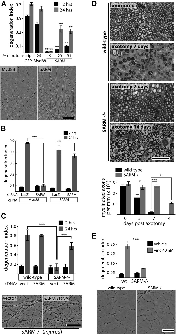
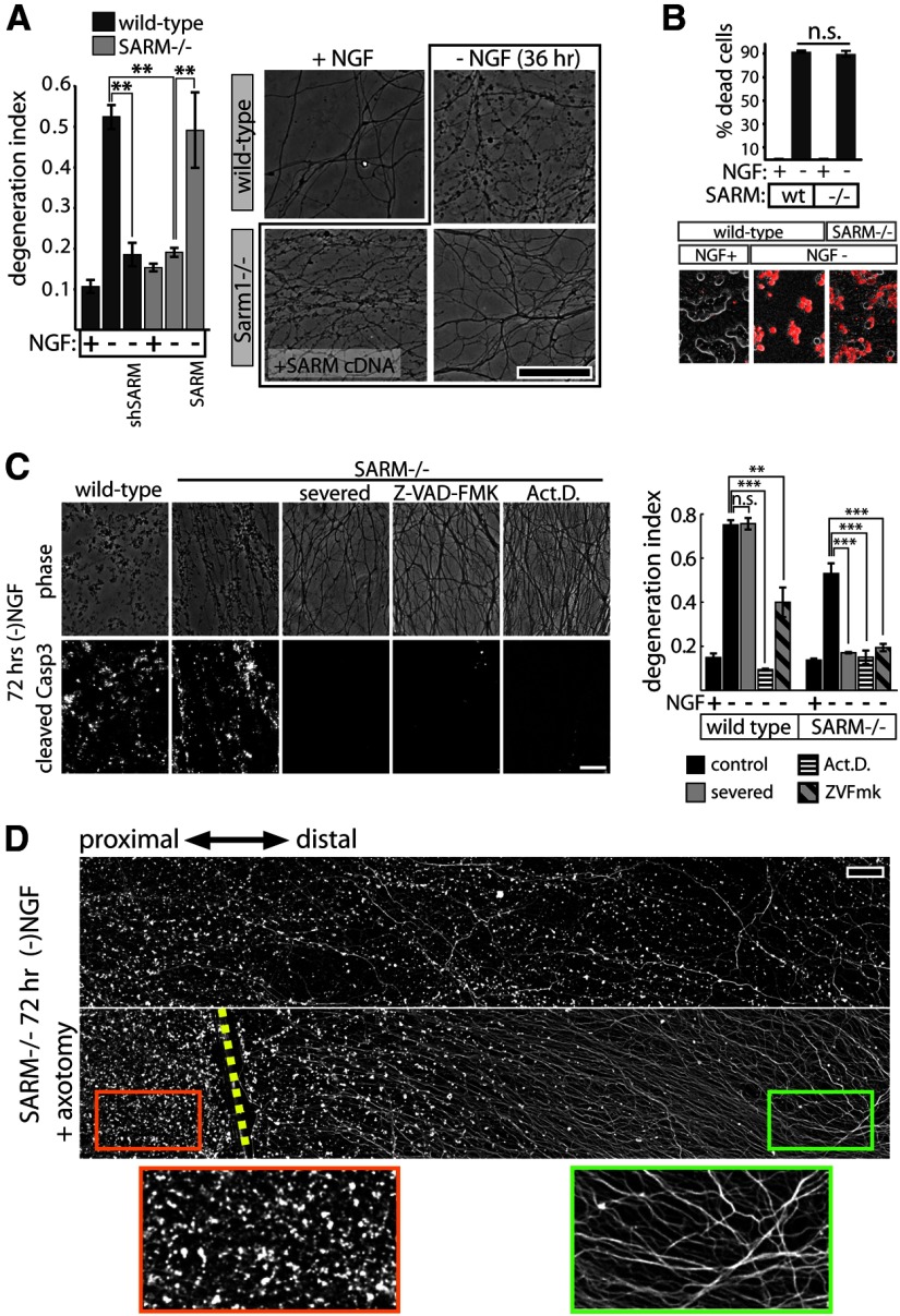
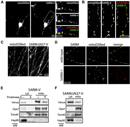
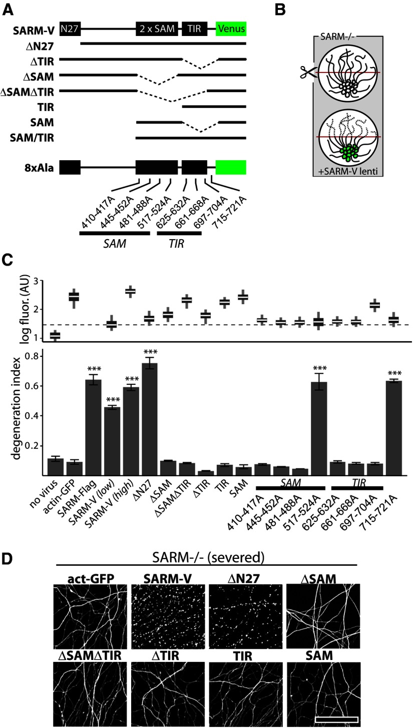
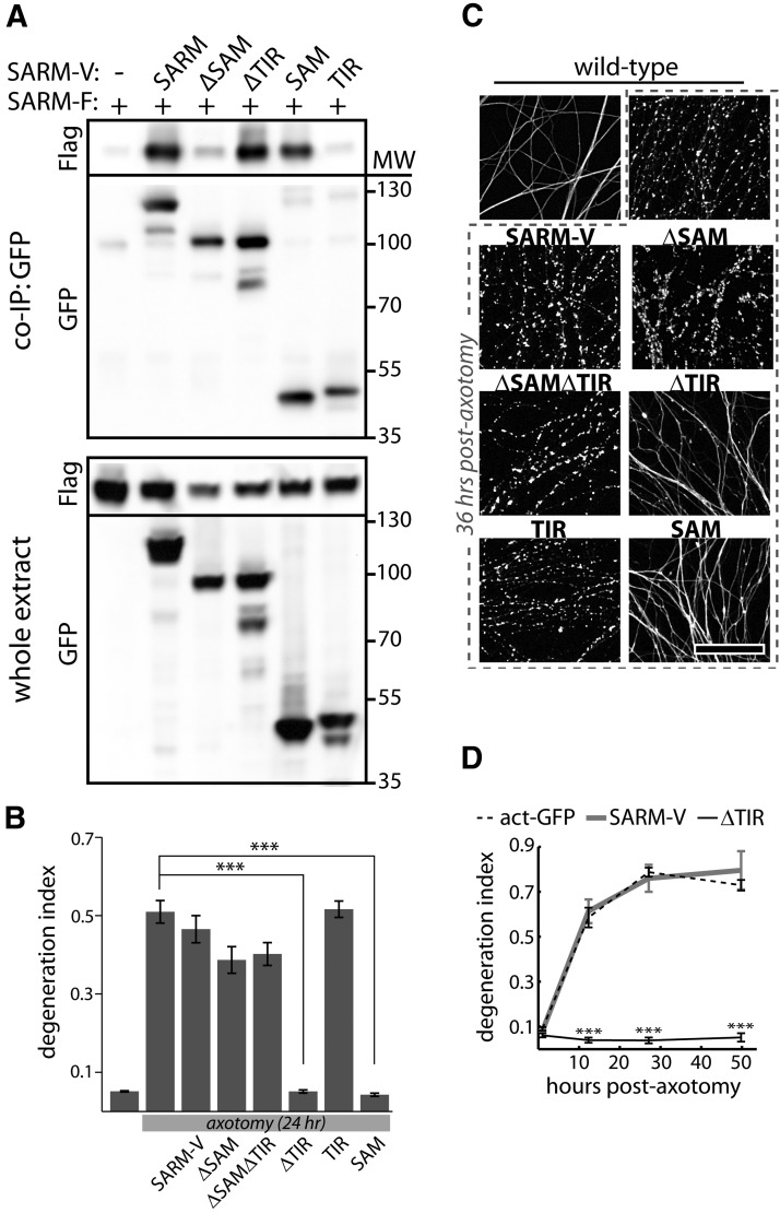
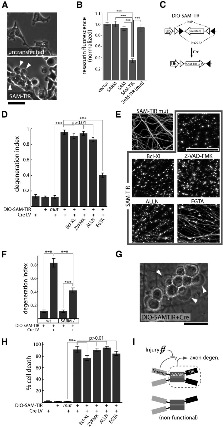
Axon degeneration is an evolutionarily conserved pathway that eliminates damaged or unneeded axons. Manipulation of this poorly understood pathway may allow treatment of a wide range of neurological disorders. In an RNAi-based screen performed in cultured mouse DRG neurons, we observed strong suppression of injury-induced axon degeneration upon knockdown of Sarm1 [SARM (sterile α-motif-containing and armadillo-motif containing protein)]. We find that a SARM-dependent degeneration program is engaged by disparate neuronal insults: SARM ablation blocks axon degeneration induced by axotomy or vincristine treatment, while SARM acts in parallel with a soma-derived caspase-dependent pathway following trophic withdrawal. SARM is a multidomain protein that associates with neuronal mitochondria. Deletion of the N-terminal mitochondrial localization sequence disrupts SARM mitochondrial localization in neurons but does not alter its ability to promote axon degeneration. In contrast, mutation of either the SAM (sterile α motif) or TIR (Toll-interleukin-1 receptor) domains abolishes the ability of SARM to promote axonal degeneration, while a SARM mutant containing only these domains elicits axon degeneration and nonapoptotic neuronal death even in the absence of injury. Protein-protein interaction studies demonstrate that the SAM domains are necessary and sufficient to mediate SARM-SARM binding. SARM mutants lacking a TIR domain bind full-length SARM and exhibit strong dominant-negative activity. These results indicate that SARM plays an integral role in the dismantling of injured axons and support a model in which SAM-mediated multimerization is necessary for TIR-dependent engagement of a downstream destruction pathway. These findings suggest that inhibitors of SAM and TIR interactions represent therapeutic candidates for blocking pathological axon loss and neuronal cell death.


Figures

Figure 1.
Figure 1.
SARM is required for injury-induced axon degeneration. A, shRNAs targeting GFP (control) or Myd88 do not suppress axotomy-induced axon degeneration while SARM knockdown with three independent shRNA vectors protects axons; protection is correlated with knockdown efficiency measured by qRT-PCR (percentage remaining transcript shown below bar graph). **p < 0.01; comparisons made to shRNA sequence targeting GFP at each time point. Images of axons treated with Myd88 or SARM shRNA at 24 h postaxotomy are shown. B, Axotomy-induced axon degeneration is blocked by SARM shRNA but not control (LacZ) shRNA. Expression of human SARM cDNA does not cause direct axon degeneration (see 2 h postaxotomy) but restores normal injury-induced axon degeneration. C, Axons of wild-type DRG neurons undergo degeneration by 24 h postaxotomy while SARM−/− neurons do not. Expression of human SARM cDNA restores normal injury-induced degeneration. D, Toluidine blue-stained sciatic nerve cross sections distal to nerve transection show complete axon degeneration (loss of normal myelin profiles) by 7 d postinjury in wild-type animals while SARM−/− axons show no degeneration at 7 d and only partial degeneration by 14 d postinjury. E, Treatment with vincristine (40 nm) for 24 h causes axon fragmentation in wild-type DRG neurons but not SARM−/− neurons. Representative phase-contrast images are shown. *p < 0.05; **p < 0.01; ***p < 0.001; error bars show standard error of the mean (SEM), scale bars, 50 μm.
Figure 2.
Figure 2.
SARM plays a role in axon degeneration elicited by trophic factor withdrawal. A, NGF deprivation elicits axon degeneration in wild-type neurons that is suppressed by SARM knockdown (shSARM). SARM−/− neurons show suppressed axon degeneration that is restored by expression of SARM. Representative phase-contrast images are shown at the right. B, SARM ablation does not affect apoptosis of DRG neurons measured after 24 h of NGF deprivation using ethidium homodimer (EH) exclusion assay. Top, Quantification of EH-positive cells shows no difference in cell death between genotypes. Bottom, Representative images of EH positivity (red stain). C, Following 72 h trophic factor withdrawal, SARM−/− axons exhibit fragmentation and cleaved caspase-3 (Casp3) immunoreactivity comparable to wild-type axons. Axon degeneration and Casp3 are blocked by severing the axons, treating with caspase inhibitor Z-VAD-FMK (10 μg/ml), or the transcriptional inhibitor actinomycin D (Act.D.; 1 μg/ml). Axon degeneration quantification is shown in the bar graph (right). D, Fluorescence (α-tubulin stain) montages showing axotomy-induced axon protection in SARM−/− DRGs deprived of NGF for 72 h. Cut site is indicated by yellow dashed line. **p < 0.01; ***p < 0.001; error bars = SEM, scale bars, 50 μm.
Figure 3.
Figure 3.
SARM associates with neuronal mitochondria. A, A SARM-Venus fusion protein (SARM-V) expressed in cortical neurons colocalizes with mitochondria-targeted DSRed protein (mitoDSRed) in both soma and neurites. B, DRG axons expressing mitoDSRed and SARM-V show mitochondrial colocalization. C, Immunostaining of endogenous SARM in DRG axons shows colocalization with mitoDSRed. A lack of immunoreactivity in SARM−/− axons (bottom left) verifies the specificity of the antibody used. D, SARMΔN27 mutant is localized diffusely in DRG axons and does not colocalize with mitoDSRed protein. E, SARM-V cofractionates with mitochondrial resident proteins Opa1 and Tom40. Treatment of mitochondrial and cytosolic fractions with proteinase K leads to proteolysis of SARM but not Opa1 or Tom40, indicating SARM-V resides outside the mitochondria. F, SARM lacking residues 2–27 (SARMΔN27-V) predominantly fractionates with cytosolic protein (ex.Hsp90). Scale bars, 10 μm.
Figure 4.
Figure 4.
SAM and TIR domains of SARM are required to mediate injury-induced axon degeneration. A, Diagram of Venus-tagged full-length SARM (SARM-V) and various mutants analyzed. Dotted lines indicate deleted regions. N27, Residues 1–27. Eight separate alanine replacement mutants (8×Ala) contain eight Ala residues in place of the indicated SARM residues. B, Genetic rescue experiment schematic: SARM−/− axons do not degenerate upon injury whereas axons of SARM−/− neurons infected with SARM lentivirus are competent to undergo injury-induced degeneration. C, Bottom, Axons of SARM−/− DRG neurons expressing actin-GFP (control) do not degenerate by 36 h postaxotomy; normal axon degeneration is restored by expression of Flag-tagged SARM (SARM-Flag), SARM-V, and ΔN27 constructs but not ΔSAM, ΔSAMΔTIR, ΔTIR, TIR, or SAM constructs. Three of four 8×Ala mutants disrupting the SAM domains and three of three disrupting the TIR domain abolished axon degeneration function of SARM while a C-terminal 8×Ala replacement outside the TIR domain did not block function. These results are also summarized in Tables 1 and 2. Top, Box plot depicts log fluorescence intensity measured from >100 cells per group (see Materials and Methods); wide boxes denote lower and upper quartiles, white lines denote median values, and whiskers extend to upper and lower 95th percentiles. Median fluorescence of all mutants exceeds that of a functional low-virus control (SARM-V low; dashed line). ***p < 0.001; one-way ANOVA with Tukey–Kramer post-test. D, Representative α-tubulin-stained axon images for deletion mutants shown in B. Error bars = SEM, scale bars, 50 μm.
Figure 5.
Figure 5.
SARM forms SAM-mediated complexes that require multiple TIR domains to promote axon degeneration. A, Coimmunoprecipitation of full-length Flag-tagged SARM (SARM-F) with Venus-tagged SARM and deletion mutants. Full-length SARM-V and mutants containing SAM domains (ΔTIR, SAM) interact strongly with full-length SARM (top, Flag blot), whereas little interaction is observed for mutants lacking SAM domains (ΔSAM, TIR). B, Axons of wild-type DRG neurons degenerate within 24 h postaxotomy. Degeneration is unaffected by expression of SARM-V, ΔSAM, ΔSAMΔTIR, or TIR, but is blocked by SARM mutants that lack a TIR domain but contain intact SAM domains (ΔTIR, SAM). C, Representative images of α-tubulin-stained axons from experiment shown in B. D, Time course measurement of axon degeneration following axotomy. While SARM-V expression does not affect the rate of axon degeneration, expression of SARM lacking a TIR domain (ΔTIR) completely blocks axon degeneration for ≥2 d. ***p < 0.001; error bars = SEM, scale bar, 50 μm.
Figure 6.
Figure 6.
SAM-TIR expression induces axonal degeneration and nonapoptotic neuronal death. A, SAM-TIR expression induces pronounced blebbing (arrowheads) in HEK293 cells at 24 h post-transfection. B, SAM-TIR overexpression in HEK293 cells leads to a loss of cell viability measured by the metabolic resazurin assay at 36 h post-transfection. Cell viability is unaffected by expression of full-length SARM, SAM, or mutated SAM-TIR bearing an 8×Ala replacement mutation in the TIR domain that blocks SARM function in axon degeneration (SAM-TIR mut = [697-704A]). ***p < 0.001 unpaired t test with Bonferroni correction. C, Lentiviral packaging of SAM-TIR is made possible by a conditionally expressed DIO SAM-TIR: a lentivirus construct containing inverted SAM-TIR-GFP flanked by loxp and lox2722 sites is conditionally expressed in cells only upon Cre-dependent recombination. Ub, Human ubiquitin C promoter. D, SAM-TIR expression (DIO-SAM-TIR lentivirus plus Cre lentivirus) induces axon degeneration that is not blocked by the apoptosis-inhibiting protein Bcl-XL, pan-caspase inhibitor Z-VAD-FMK (ZVFMK; 10 μg/ml), or calpain inhibitor ALLN (25 μm). Chelation of extracellular Ca ions with EGTA (2.5 mm) provides partial suppression of axon degeneration. E, Representative α-tubulin-stained axons from experiment shown in D. F, SAM-TIR expression induces axon degeneration in both wild-type and SARM−/− axons measured 3 d postinfection; ***p < 0.001 unpaired t tests with Bonferroni correction. G, SAM-TIR expression induces large translucent neuronal swellings evident at 2 d postinfection. H, SAM-TIR expression induces neuronal death measured by ethidium homodimer staining at 3 d postinfection. Cell death is not blocked by the apoptosis-inhibiting protein Bcl-XL, pan-caspase inhibitor Z-VAD-FMK (ZVFMK; 10 μg/ml), calpain inhibitor ALLN (25 μm), or chelation of extracellular Ca ions with EGTA (2.5 mm). ***p < 0.001 unpaired t tests with Bonferroni correction. I, Proposed model, SARM forms SAM-mediated complexes that promote axon degeneration via intracomplex TIR interactions. Top, In uninjured cells, the N terminus of SARM suppresses the degeneration-promoting C terminus. Injury leads to release of this inhibition. Bottom, Incorporation of TIR-less molecules into SARM complexes renders them nonfunctional. Scale bars, 50 μm, error bars = SEM.

Similar articles

Cited by

References

    1. Akhouayri I, Turc C, Royet J, Charroux B. Toll-8/Tollo negatively regulates antimicrobial response in the Drosophila respiratory epithelium. PLoS Pathogens. 2011;7:e1002319. doi: 10.1371/journal.ppat.1002319. - DOI - PMC - PubMed
    1. Araki T, Sasaki Y, Milbrandt J. Increased nuclear NAD biosynthesis and SIRT1 activation prevent axonal degeneration. Science. 2004;305:1010–1013. doi: 10.1126/science.1098014. - DOI - PubMed
    1. Babetto E, Beirowski B, Russler EV, Milbrandt J, Diantonio A. The Phr1 ubiquitin ligase promotes injury-induced axon self-destruction. Cell Rep. 2013;3:1422–1429. doi: 10.1016/j.celrep.2013.04.013. - DOI - PMC - PubMed
    1. Barrientos SA, Martinez NW, Yoo S, Jara JS, Zamorano S, Hetz C, Twiss JL, Alvarez J, Court FA. Axonal degeneration is mediated by the mitochondrial permeability transition pore. J Neurosci. 2011;31:966–978. doi: 10.1523/JNEUROSCI.4065-10.2011. - DOI - PMC - PubMed
    1. Bhattacharya MR, Gerdts J, Naylor SA, Royse EX, Ebstein SY, Sasaki Y, Milbrandt J, DiAntonio A. A model of toxic neuropathy in Drosophila reveals a role for MORN4 in promoting axonal degeneration. J Neurosci. 2012;32:5054–5061. doi: 10.1523/JNEUROSCI.4951-11.2012. - DOI - PMC - PubMed

Publication types

MeSH terms