. 2013 Sep 3;110(36):E3445-54.
doi: 10.1073/pnas.1303002110. Epub 2013 Aug 14.

Stapled α-helical peptide drug development: a potent dual inhibitor of MDM2 and MDMX for p53-dependent cancer therapy

Affiliations

Stapled α-helical peptide drug development: a potent dual inhibitor of MDM2 and MDMX for p53-dependent cancer therapy

Yong S Chang et al. Proc Natl Acad Sci U S A. .

Abstract

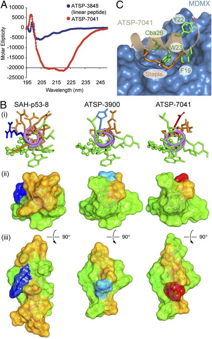
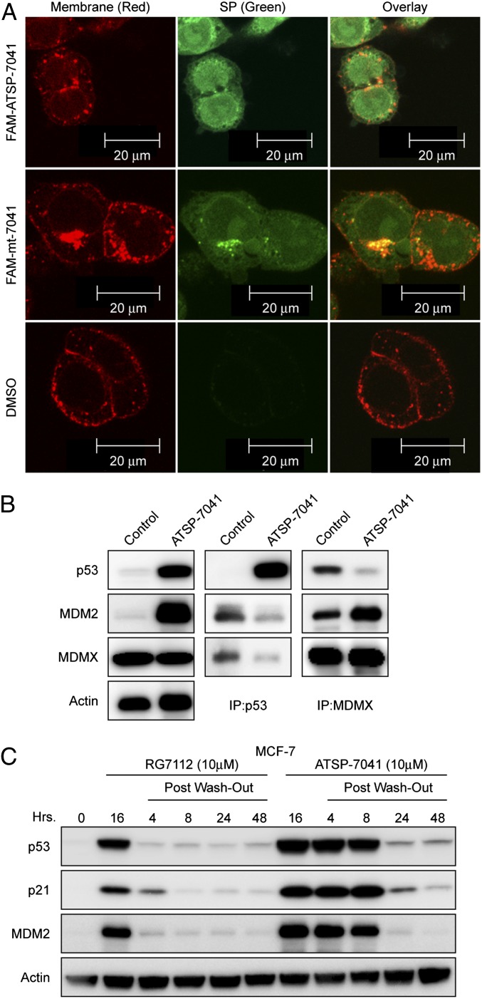
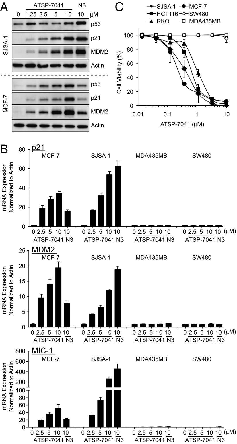
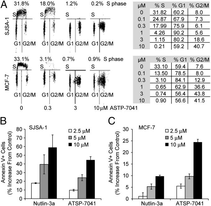
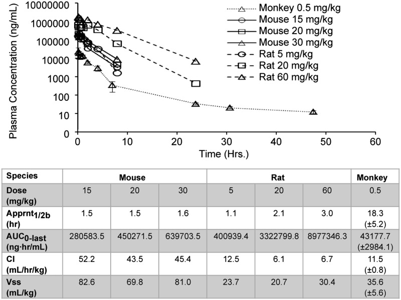
Stapled α-helical peptides have emerged as a promising new modality for a wide range of therapeutic targets. Here, we report a potent and selective dual inhibitor of MDM2 and MDMX, ATSP-7041, which effectively activates the p53 pathway in tumors in vitro and in vivo. Specifically, ATSP-7041 binds both MDM2 and MDMX with nanomolar affinities, shows submicromolar cellular activities in cancer cell lines in the presence of serum, and demonstrates highly specific, on-target mechanism of action. A high resolution (1.7-Å) X-ray crystal structure reveals its molecular interactions with the target protein MDMX, including multiple contacts with key amino acids as well as a role for the hydrocarbon staple itself in target engagement. Most importantly, ATSP-7041 demonstrates robust p53-dependent tumor growth suppression in MDM2/MDMX-overexpressing xenograft cancer models, with a high correlation to on-target pharmacodynamic activity, and possesses favorable pharmacokinetic and tissue distribution properties. Overall, ATSP-7041 demonstrates in vitro and in vivo proof-of-concept that stapled peptides can be developed as therapeutically relevant inhibitors of protein-protein interaction and may offer a viable modality for cancer therapy.


Conflict of interest statement

Conflict of interest statement: All authors are employees of either Aileron Therapeutics, Inc. or Hoffmann-La Roche, Inc. and, in each case, are shareholders of the company for which they work.

Figures

Fig. 1.
Fig. 1.
The chemical structure of ATSP-7041 is shown highlighting the R8 (blue) and S5 (red) amino acids in the stapled peptide following ring-closing metathesis (and loss of ethylene). Note that each R8 and S5 is α-methylated and that the all-hydrocarbon linker consists of 11 carbon atoms. (A) Sequences of a series of stapled peptides and their binding properties to MDM2 and MDMX. Lead optimization of the linear phage display peptide (pDI) resulted in the highly potent lead stapled peptide ATSP-7041. ATSP-7041 exhibits high-affinity binding to both MDM2 and MDMX versus the small-molecule Nutlin-3a. The Phe19 to Ala19 mutant negative control analog (ATSP-7342) is essentially devoid of binding to MDM2 and MDMX as expected. (B) Biacore studies confirmed the high affinity of ATSP-7041 to MDM2 and MDMX and also reveal its fast on-rate but rather slow off-rate for target binding. (C) ATSP-7041 exhibits high potency in SJSA-1 cell viability assay in the presence of increasing serum in contrast to that of stapled peptides SAH-p53-8 and the pDI analog ATSP-3900.
Fig. 2.
Fig. 2.
Conformational and 3D-structural properties of ATSP-7041. (A) Comparative CD spectroscopy of pDI and stapled peptide ATSP-7041in pH 7 buffer illustrates their intrinsic α−helical properties. (B) The α−helical wheel projections (3D) of ATSP-7041 versus SAH-p53-8 and the stapled pDI analog ATSP-3900 show the extended hydrophobic surface contributing to the enhance amphipathicity of ATSP-7041. (i) The three stapled peptides are represented as fully α-helical peptides with the viewer facing the long axis of the helix by the N terminus. Side chains are represented in stick style whereas the olefin staple is in ball and stick style. Polar residues (Asn, Gln, Ser, and Thr) are colored orange; green is for hydrophobic, olefin staple, and aromatic residues (Ala, Val, Leu, Ile, Pro, Cba, Phe, Tyr, and Trp), blue for basic residues (Lys and Arg), red for acidic residues (Asp and Glu), and light blue for His. (ii) Solvent exposed surfaces representation of the three stapled peptides with the same view angle as in ref. and the same coloring. (iii) Other view of the solvent exposed surfaces, after vertical rotation of 90°. (C) A high-resolution X-ray structure of ATSP-7041 bound to MDMX delineates its intermolecular contacts with the target protein. The side chain is colored in green: Phe19, Trp23, Cba26, and Tyr22 are the main interacting residues responsible for the high binding affinity whereas the staple olefin, also contributing to the binding affinity, is colored in orange (see Results for detailed discussion).
Fig. 3.
Fig. 3.
ATSP-7041 penetrates cell membranes and disrupts p53-MDM2 and p53-MDMX binding. (A) A fluorescent FAM was linked via a β-alanine spacer to the N terminus of ATSP-7041. HCT116 cells were seeded at 60,000 cells per well and incubated with 20 μM stapled peptides or DMSO control for 4.5 h. Cells were imaged using an LSM 510 Zeiss Axiovert 200M (v4.0) confocal microscope using the 63× oil lens. The equipotent FAM-labeled ATSP-7041 showed a diffused intracellular localization, demonstrating efficient cellular penetration. Similarly the FAM-mt-7041 containing single F19 to A19 modification that exhibits no binding or cell activity also showed efficient cell penetration, confirming that its inactivity is due to lack of binding to the target proteins. (B) ATSP-7041 inhibits p53-MDM2 and p53-MDMX binding in cancer cells. MCF-7 cells were incubated with 10 μM ATSP-7041 for 4 h, and the levels of p53, MDM2, and MDMX were determined in protein complexes immunoprecipitated with anti-MDMX or anti-p53 antibodies by Western blotting. ATSP-7041 induces a dose-dependent decrease in the level of both MDM2 and MDMX associated with p53 in comparison with the vehicle control (DMSO). Similarly, IP of MDMX from ATSP-7041 dosed MCF-7 cells shows a significant dose-dependent decrease in the level of p53 in the complex in comparison with the vehicle control (DMSO). ATSP-7041 does not inhibit the complex between MDM2 and MDMX. (C) ATSP-7041induced a more durable effect on p53 signaling than small-molecule, MDM2-selective inhibitors. Induction of p53, p21, and MDM2 was unchanged up to 4 h after drug removal and continued to be elevated above the control up to 48 h in response to ATSP-7041treatment. In contrast, RG7112 lost most of its inhibitory effect at 4 h after media replacement and maintained p53 and target protein levels only slightly above basal levels.
Fig. 4.
Fig. 4.
ATSP-7041 activates p53 signaling in cancer cells in p53WT cell lines. (A) ATSP-7041 stabilizes p53 and elevates protein levels of p53 targets p21 and MDM2. Log-phase SJSA-1 and MCF-7 cells were incubated with 1.25, 2.5, 5.0, or 10 μM ATSP-7041 or 10 μM Nutlin-3a for 24 h, and cell lysates were analyzed by Western blotting. (B) ATSP-7041 shows dose-dependent induction of p53 target genes in p53WT but not in mutant p53 cell lines. Exponentially growing p53WT (SJSA-1, MCF-7) and p53 mutant (SW480, MDA-MD-435) cancer cell lines were incubated with 2.5, 5, or 10 μM ATSP-7041 or 10 μM Nutlin-3a for 24 h, and mRNA levels of p53 targets, p21, MDM2, and MIC-1 were measured by quantitative PCR and expressed as fold increase. (C) Viability of four p53WT (SJSA-1, MCF-7, HCT-116, and RKO) and two mutant p53 (SW480 and MDA-MB-435) cancer cell lines was determined after incubation with ATSP-7041 and expressed as percentage of controls ± SD.
Fig. 5.
Fig. 5.
ATSP-7041 activates main p53 cellular functions in cancer cells. (A) ATSP-7041 arrests cell-cycle progression in cancer cells with p53WT. Exponentially growing SJSA-1 and MCF-7 cells were incubated with 0.3, 3, or 10 μM ATSP-7041for 24 h, and cell cycle distribution was determined by BrdU labeling and cell-cycle analysis. Numbers indicate the percentage of S-phase cells. (B) Apoptotic response to ATSP-7041 and Nutlin-3a in SJSA-1 MDM-2 sensitive cell line. SJSA-1 cells were exposed to ATSP-7041(2.5, 5, or 10 μM) and Nutlin-3a (2.5, 5, or 10 μM) for 48 h, and the percentage of apoptotic cells (±SD) was determined by the Annexin V assay. (C) Apoptotic response to ATSP-7041 and Nutlin-3a in the MCF-7 line. MCF-7 cells were exposed to ATSP-7041 (2.5, 5, or 10 μM) and Nutlin-3a (2.5, 5, or 10 μM) for 48 h, and apoptosis was determined by the Annexin V assay.
Fig. 6.
Fig. 6.
ATSP-7041 suppresses tumor growth in vivo of multiple human xenograft models and increases expression of p21. (A) ATSP-7041 induced statistically significant tumor growth inhibition for both qd and qod dosing schedules against the MDM2 amplified osteosarcoma xenograft model SJSA-1. Tumor volumes were calipered throughout the study, and data were plotted as mean ± SEM. *P < 0.05 vs. vehicle control. (B) Treatments resulted in dose-dependent and statistically significant tumor growth inhibition against the MDMX overexpressing MCF-7 human breast cancer xenograft model. Mice (n = 10 per group) bearing established s.c. xenografts received vehicle or 20 or 30 mg/kg ATSP-7041 solution i.v., qod, or 50 or 100 mg/kg of a p.o. suspension of RG7112 daily. *P < 0.05 vs. vehicle control. (C) ATSP-7041 induced greater p21 expression than the MDM2 selective small-molecule inhibitor RG7112. Tumors were removed from mice 4, 8, or 24 h after the last dose of either vehicle or ATSP-7041 at 20 or 30 mg/kg, and p21 mRNA levels were determined by quantitative RT-PCR. Induction of p21 was observed for tumors in response to ATSP-7041 treatment, with a robust 12- to 19-fold increase in p21 8 h post dose. In contrast, for RG7112, only modest induction of p21 was observed at all doses and time points.
Fig. 7.
Fig. 7.
ATSP-7041 exhibits favorable DMPK properties. Shown are mean plasma pharmacokinetic profiles of ATSP-7041 via i.v. administration in female nude mice, Sprague–Dawley rat, and cynomolgus monkeys. Female nude mice are represented by solid lines (sparse sampling with n of 2 at each time point): 15 mg/kg (circles), 20 mg/kg (squares), and 30 mg/kg (triangles). Sprague–Dawley rats are represented by dashed lines (serial sampling with n = 2 at each time point): 5 mg/kg (circles), 20 mg/kg (squares), and 60 mg/kg (triangles). Cynomolgus monkeys are represented by dotted lines (serial sampling with n = 3 at each time point): 0.5 mg/kg (triangles).
Fig. 8.
Fig. 8.
ATSP-7041 exhibits favorable tissue distribution properties. Mean representative whole body autoradioluminograms showing tissue distribution of [3H]ATSP-7041–derived radioactivity at 0.5 (A) and 6 h (B) after an i.v. dose of [3H]ATSP-7041 at 5 mg/kg to male Long–Evans rats. Highly vascularized tissues, including essential target tissues for the treatment of solid and hematologic tumors, showed the greatest tissue-to-plasma AUC0-t ratios.

Comment in

References

    1. Cheok CF, Verma CS, Baselga J, Lane DP. Translating p53 into the clinic. Nat Rev Clin Oncol. 2011;8(1):25–37. - PubMed
    1. Vogelstein B, Lane D, Levine AJ. Surfing the p53 network. Nature. 2000;408(6810):307–310. - PubMed
    1. Bernal F, et al. A stapled p53 helix overcomes HDMX-mediated suppression of p53. Cancer Cell. 2010;18(5):411–422. - PMC - PubMed
    1. Marine JC. MDM2 and MDMX in cancer and development. Curr Top Dev Biol. 2011;94:45–75. - PubMed
    1. Migliorini D, et al. Mdm4 (Mdmx) regulates p53-induced growth arrest and neuronal cell death during early embryonic mouse development. Mol Cell Biol. 2002;22(15):5527–5538. - PMC - PubMed

MeSH terms