. 2013 Sep;140(18):3882-91.
doi: 10.1242/dev.095687. Epub 2013 Aug 14.

FGF signaling regulates Wnt ligand expression to control vulval cell lineage polarity in C. elegans

Affiliations

FGF signaling regulates Wnt ligand expression to control vulval cell lineage polarity in C. elegans

Paul J Minor et al. Development. 2013 Sep.

Abstract

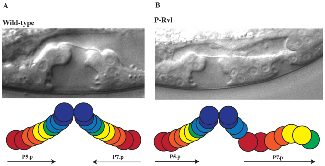
The interpretation of extracellular cues leading to the polarization of intracellular components and asymmetric cell divisions is a fundamental part of metazoan organogenesis. The Caenorhabditis elegans vulva, with its invariant cell lineage and interaction of multiple cell signaling pathways, provides an excellent model for the study of cell polarity within an organized epithelial tissue. Here, we show that the fibroblast growth factor (FGF) pathway acts in concert with the Frizzled homolog LIN-17 to influence the localization of SYS-1, a component of the Wnt/β-catenin asymmetry pathway, indirectly through the regulation of cwn-1. The source of the FGF ligand is the primary vulval precursor cell (VPC) P6.p, which controls the orientation of the neighboring secondary VPC P7.p by signaling through the sex myoblasts (SMs), activating the FGF pathway. The Wnt CWN-1 is expressed in the posterior body wall muscle of the worm as well as in the SMs, making it the only Wnt expressed on the posterior and anterior sides of P7.p at the time of the polarity decision. Both sources of cwn-1 act instructively to influence P7.p polarity in the direction of the highest Wnt signal. Using single molecule fluorescence in situ hybridization, we show that the FGF pathway regulates the expression of cwn-1 in the SMs. These results demonstrate an interaction between FGF and Wnt in C. elegans development and vulval cell lineage polarity, and highlight the promiscuous nature of Wnts and the importance of Wnt gradient directionality within C. elegans.

Keywords: C. elegans; Cell polarity; FGF; Vulval development; Wnt.


Figures

Fig. 1.
Fig. 1.
C. elegans vulval development. (A) Schematic of vulval induction illustrating sources of EGF, Notch and Wnt. AC, anchor cell. (B) Lineage trees of VPC progeny: P5.p, secondary (2°) fate on the left; P6.p, primary (1°) fate in center; and P7.p, secondary 2° fate on the right. The progeny of each cell is color coded: A cells, red; B cells (B1 and B2), orange; C cells, yellow; D cells, green; E cells, light blue; F cells, dark blue. (C) Final conformation of vulval cells shown as a cartoon and Nomarski image in mid-L4 stage. Mirror symmetry is noted about the vulval center. Proximal daughter cells of P5.p and P7.p join the daughters of P6.p in forming the vulval lumen, whereas the distal most daughters of P5.p and P7.p adhere to the ventral epidermis. (D) The default polarity of P5.p and P7.p is random in the absence of all Wnts. (E) EGL-20 is expressed in the tail (green circles) and establishes ground polarity in which both P5.p and P7.p face the posterior as a result of asymmetric localization of SYS-1, LIT-1 and WRM-1 to the posterior daughter of P7.p and POP-1 to the anterior daughter. (F) LIN-44 and MOM-2 are expressed in the anchor cell (yellow circle) resulting in refined polarity in which P5.p and P7.p both face towards the center as a result of asymmetric localization of SYS-1, LIT-1 and WRM-1 to the anterior daughter cell of P7.p and POP-1 to the posterior daughter cell.
Fig. 2.
Fig. 2.
Wild-type vulva versus posterior-reversed vulval lineage vulva. (A) Wild-type vulva formed from 22 progeny of three VPCs: P5.p, P6.p and P7.p. The progeny of P5.p and P7.p form mirror symmetry about the vulval center. (B) Posterior-reversed vulval lineage: the daughter cells of P7.p mimic those of P5.p. Both images taken with sem-5(n1779) background. Color key as described for Fig. 1.
Fig. 3.
Fig. 3.
egl-17::gfp expression in P6.p. egl-17 is activated by EGF signaling and is expressed in P6.p. Expression of egl-17 is used as a marker for vulval induction.
Fig. 4.
Fig. 4.
Subcellular localization of VNS::SYS-1. The localization pattern of VNS::SYS-1 in P7.p daughter cells. The resulting pattern was classified by eye into three categories: SYS-1 enriched in the anterior daughter (P7.pa >P7.pp), SYS-1 present at similar levels in both daughters (P7.pa=P7.pp), and SYS-1 enriched in the anterior daughter (P7.pa <P7.pp). A representative image of each scenario is shown. Graph shows percentage of animals with the pattern of SYS-1 localization indicated.
Fig. 5.
Fig. 5.
P6.p influences the polarity of P7.p. (A) Prior to induction, the anchor cell (AC) is directly dorsal to P6.p. (B) During induction, the anchor cell produces LIN-3, which is supplied to P5-7.p. Induction activates egl-17, illustrated in blue, within P6.p. (C) P6.p is ablated at the start of induction, but prior to the VPC polarity choice, leaving only P5.p and P7.p, the secondary VPCs. (D) Wild-type orientation of a worm with P6.p ablated. (E). P-Rvl orientation of a worm with P6.p ablated. (F) Ablating P6.p enhances the phenotype of lin-18(e620).
Fig. 6.
Fig. 6.
The sex myoblasts influence the polarity of P7.p. (A) The sex myoblasts arise from the M cell as shown in the cell lineage. The sex myoblasts are born 13 hours post-hatch and migrate anteriorly until they reach their final position, flanking the center of the gonad. The polarity decision of P7.p is made after the sex myoblasts have reached their final position and prior to their first division. The M cell/sex myoblasts are shown in purple, the gonad blue and the VPCs green. (B) The M cell lineage shown in a sem-2(n1343) background. The SMs do not form in this background, but instead become posterior body wall muscle, marked in red and with an asterisk.
Fig. 7.
Fig. 7.
FGF signaling regulates cwn-1 expression in the SMs. (A) smFISH analysis of the cwn-1 transcript in a wild-type worm at the time of the polarity decision. cwn-1 is predominantly expressed in the posterior body wall muscle (BWM) and in the M cell/SM lineage. (B) A histogram quantifying the number of cwn-1 transcripts per SM in wild-type (blue bars) and egl-15(n484) (red bars) backgrounds.
Fig. 8.
Fig. 8.
The role of Wnt signaling on P7.p (A) The anchor cell (blue; AC) releases LIN-3 (red arrow), inducing the VPCs (green). Induction triggers the expression of the FGF ligand EGL-17 (yellow arrow) in P6.p, which activates the FGF pathway located in the SMs (purple). The FGF pathway regulates the expression of cwn-1 in the SMs. The SMs are the anterior source of CWN-1 for P7.p whereas the posterior body wall muscle serves as the posterior source. All Wnt signals are depicted with an orange arrow. lin-44 and mom-2, both expressed anterior to P7.p, express in the anchor cell whereas egl-20 expresses in the tail, the posterior side of P7.p. (B) CWN-1 from the SMs and LIN-44 act through LIN-17, and MOM-2 acts through LIN-18. All three ligands act to drive SYS-1 localization to the anterior daughter of P7.p. Posterior-expressed CWN-1 and EGL-20 act through CAM-1 to drive SYS-1 localization to the posterior daughter of P7.p. Genetic data indicate that EGL-20 possibly acts through another, unknown receptor. (C-E) Examples of how mutations drive phenotypic output. In lin-17(n671); lin-18(e620), all anterior receptors are eliminated resulting in a 100% P-Rvl phenotype. Mutations in cwn-1 eliminate it from both sides of the pathway whereas sem-2(n1343), owing to a genetic ablation of the SMs, eliminates only the anterior source of cwn-1.

References

    1. Bargmann C. I., Avery L. (1995). Laser killing of cells in Caenorhabditis elegans. Methods Cell Biol. 48, 225–250 - PMC - PubMed
    1. Branda C. S., Stern M. J. (2000). Mechanisms controlling sex myoblast migration in Caenorhabditis elegans hermaphrodites. Dev. Biol. 226, 137–151 - PubMed
    1. Brenner S. (1974). The genetics of Caenorhabditis elegans. Genetics 77, 71–94 - PMC - PubMed
    1. Burdine R. D., Chen E. B., Kwok S. F., Stern M. J. (1997). egl-17 encodes an invertebrate fibroblast growth factor family member required specifically for sex myoblast migration in Caenorhabditis elegans. Proc. Natl. Acad. Sci. USA 94, 2433–2437 - PMC - PubMed
    1. Burdine R. D., Branda C. S., Stern M. J. (1998). EGL-17(FGF) expression coordinates the attraction of the migrating sex myoblasts with vulval induction in C. elegans. Development 125, 1083–1093 - PubMed

Publication types

MeSH terms