. 2013 Sep 27;288(39):27764-76.
doi: 10.1074/jbc.M113.476358. Epub 2013 Aug 14.

Prefoldin plays a role as a clearance factor in preventing proteasome inhibitor-induced protein aggregation

Affiliations

Prefoldin plays a role as a clearance factor in preventing proteasome inhibitor-induced protein aggregation

Akira Abe et al. J Biol Chem. .

Abstract

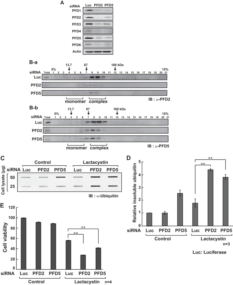
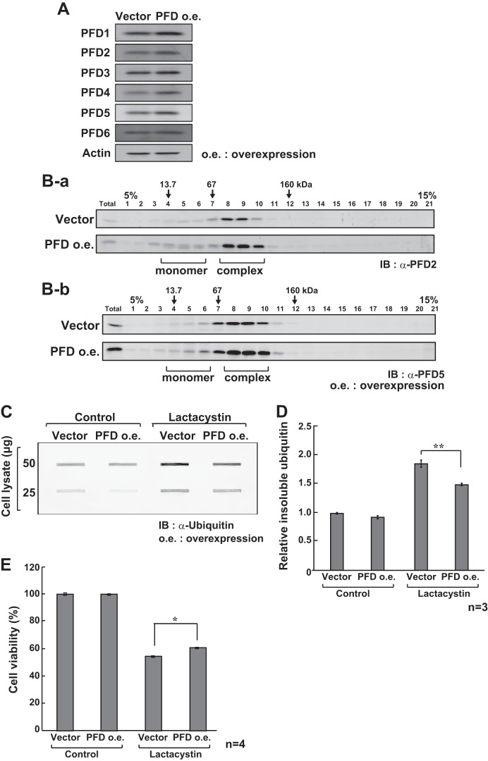
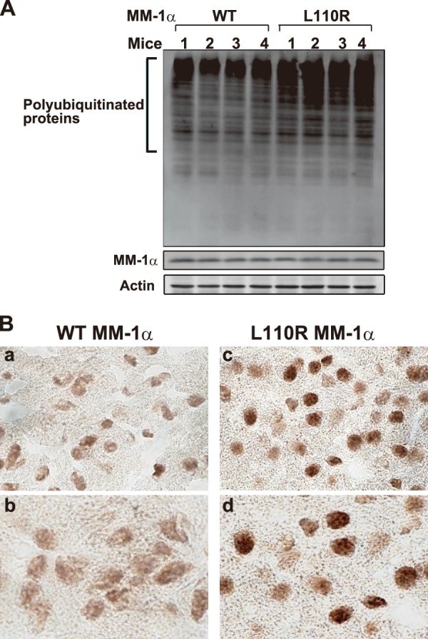
Prefoldin is a molecular chaperone composed of six subunits, PFD1-6, and prevents misfolding of newly synthesized nascent polypeptides. Although it is predicted that prefoldin, like other chaperones, modulates protein aggregation, the precise function of prefoldin against protein aggregation under physiological conditions has never been elucidated. In this study, we first established an anti-prefoldin monoclonal antibody that recognizes the prefoldin complex but not its subunits. Using this antibody, it was found that prefoldin was localized in the cytoplasm with dots in co-localization with polyubiquitinated proteins and that the number and strength of dots were increased in cells that had been treated with lactacystin, a proteasome inhibitor, and thapsigargin, an inducer of endoplasmic reticulum stress. Knockdown of prefoldin increased the level of SDS-insoluble ubiquitinated protein and reduced cell viability in lactacystin and thapsigargin-treated cells. Opposite results were obtained in prefoldin-overexpressed cells. It has been reported that mice harboring a missense mutation L110R of MM-1α/PFD5 exhibit neurodegeneration in the cerebellum. Although the prefoldin complex containing L110R MM-1α was properly formed in vitro and in cells derived from L110R MM-1α mice, the levels of ubiquitinated proteins and cytotoxicity were higher in L110R MM-1α cells than in wild-type cells under normal conditions and were increased by lactacystin and thapsigargin treatment, and growth of L110R MM-1α cells was attenuated. Furthermore, the polyubiquitinated protein aggregation level was increased in the brains of L110R MM-1α mice. These results suggest that prefoldin plays a role in quality control against protein aggregation and that dysfunction of prefoldin is one of the causes of neurodegenerative diseases.

Keywords: Cell Death; Chaperone Chaperonin; Neurodegeneration; Prefoldin; Proteasome; Protein Aggregation.


Figures

FIGURE 1.
FIGURE 1.
Localization of prefoldin. A, six purified prefoldin subunits were denatured and then renatured as described under “Experimental Procedures.” The protein mixture was then applied onto a gel filtration column (HiLoad 16/600 Superdex 200). mAU, measurable absorbance unit. B, HeLa cells were stained with an anti-prefoldin antibody (clone 1E9), and nuclei were stained with DAPI (left and right panels, respectively). Cells were then reacted with a secondary antibody, and cell images were observed using an immunofluorescence microscope. C, HeLa cells were stained with an anti-prefoldin antibody (clone 1E9) (panels a and b). The anti-prefoldin antibody was first incubated with the cross-linked prefoldin complex (panel c) or with a mixture of six prefoldin subunits (panel d) for 1 h at 4 °C and used for immunostaining of HeLa cells. D-a, reconstituted prefoldin complex composed of 25 μg each of six recombinant prefoldin subunits was separated on a blue native gel and stained with Coomassie Brilliant Blue (CBB) (left panel). The prefoldin complex (150 μg) and 25 μg each of six recombinant prefoldin subunits were separated on blue native gels and analyzed by Western blotting with anti-prefoldin complex, anti-PFD5, and anti-PFD2 antibodies (right 3 panels). D-b, anti-prefoldin complex antibody was first reacted with the reconstituted prefoldin complex or with six prefoldin subunits for 2 h at 4 °C and used for Western blot analysis of filters, in which the prefoldin complex and six prefoldin subunits were loaded.
FIGURE 2.
FIGURE 2.
Localization of prefoldin on polyubiquitinated protein aggregates in lactacystin-treated HeLa cells. A, HeLa cells were cultured with or without 10 μm lactacystin (Lac-cys) for 24 h and stained with anti-prefoldin, anti-PFD2, and anti-PFD5 antibodies. Cell images were then obtained after cells had been reacted with the respective secondary antibodies. Control indicates cells without lactacystin treatment. B, HeLa cells were cultured with 10 μm lactacystin for 0, 8, 16, and 24 h and stained with anti-prefoldin and anti-polyubiquitin antibodies. Cell images were then obtained after cells had been reacted with the respective secondary antibodies. C, HeLa cells were cultured with or without 10 μm lactacystin for 24 h. Cells were then stained with rat anti-prefoldin and rabbit anti-ubiquitin antibodies and reacted with Alexa Fluor 488-conjugated anti-rat IgG and Alexa Fluor 594-conjugated anti-rabbit IgG antibodies, respectively. Both cell images were merged. Control indicates cells without lactacystin treatment. D, HeLa cells were treated with 10 μm lactacystin for 0, 8, 16, and 24 h. Proteins extracted from cells were treated with 1% SDS and centrifuged. SDS-soluble proteins (supernatant after centrifugation) and SDS-insoluble proteins (pellet after centrifugation) were analyzed by Western blotting with anti-polyubiquitin, anti-LC3, anti-p62, and anti-actin antibodies. E, Atg5(+/+) and Atg5(−/−) cells were cultured with or without 10 μm lactacystin for 24 h and stained with an anti-prefoldin antibody. Cell images were then obtained after cells had been reacted with an Alexa Fluor 488-conjugated anti-rat IgG antibody. Control indicates cells without lactacystin treatment.
FIGURE 3.
FIGURE 3.
Effect of prefoldin knockdown on lactacystin-induced cell toxicity. A, HeLa cells were transfected with siRNAs targeting PFD2, PFD5, and luciferase (Luc). Twenty four h after transfection, proteins extracted from transfected cells were analyzed by Western blotting with anti-prefoldin subunits and anti-actin antibodies. B, proteins extracted from HeLa cells as described in the legend of A were separated by glycerol density gradients. Proteins in each fraction were then analyzed by Western blotting with anti-PFD2 (B-a) and anti-PFD5 (B-b) antibodies as described under “Experimental Procedures.” Total indicates proteins extracted from HeLa cells. C, HeLa cells were transfected with siRNAs targeting PFD2, PFD5, and luciferase. Twenty four h after transfection, cells were treated with 10 μm lactacystin for 24 h, and proteins extracted from the cells were treated with 1% SDS, analyzed by filter trap assays, and detected with an anti-ubiquitin antibody as described under “Experimental Procedures.” IB, immunoblot. D, intensities of bands in C were quantified. Values are means ± S.D. n = 3 experiments. Significance: **, p < 0.01. E, HeLa cells were transfected with siRNAs targeting PFD2, PFD5, and luciferase. Twenty four h after transfection, cells were treated with 10 μm lactacystin for 24 h, and their viability was examined by MTT assays. Values are means ± S.D. n = 4 experiments. Significance: **, p < 0.01.
FIGURE 4.
FIGURE 4.
Effect of overexpression of prefoldin on lactacystin-induced cell toxicity. A, HeLa cells were transfected with expression vectors for six prefoldin subunits. Forty eight h after transfection, proteins extracted from transfected cells were analyzed by Western blotting with anti-prefoldin subunits and anti-actin antibodies. B, proteins extracted from HeLa cells as described in the legend of A were separated by glycerol density gradients. Proteins in each fraction were then analyzed by Western blotting with anti-PFD2 (B-a) and anti-PFD5 (B-b) antibodies as described under “Experimental Procedures.” Total indicates proteins extracted from HeLa cells. C, HeLa cells were transfected with expression vectors for six prefoldin subunits. Forty eight h after transfection, cells were treated with 10 μm lactacystin for 24 h, and proteins extracted from the cells were treated with 1% SDS, analyzed by filter trap assays, and detected with an anti-ubiquitin antibody as described under “Experimental Procedures.” IB, immunoblot. D, intensities of bands in C were quantified. Values are means ± S.D. n = 3 experiments. Significance: **, p < 0.01. E, HeLa cells were transfected with expression vectors for six prefoldin subunits. Forty eight h after transfection, cells were treated with 10 μm lactacystin for 24 h, and their viability was examined by MTT assays. Values are means ± S.D. n = 4 experiments. Significance: *, p < 0.05.
FIGURE 5.
FIGURE 5.
Effect of L110R mutation of MM-1α/PFD5 on prefoldin formation. A, five prefoldin subunits (PFD1, PFD2, PFD3, PFD4, and PFD) and wild-type or L110R MM-1α/PFD5 were expressed in and purified from E. coli. The prefoldin complex was reconstituted using these six subunits as described under “Experimental Procedures” and applied onto a gel filtration column (a). mAU, measurable absorbance unit. Proteins in fractions corresponding to the prefoldin complex were analyzed by Western blotting with anti-prefoldin subunits antibodies (b). B, proteins extracted from wild-type and L110R MM-1α cells were separated by glycerol density gradients. Proteins in each fraction were then analyzed by Western blotting with anti-PFD2 and anti-PFD5 antibodies as described under “Experimental Procedures.” Total indicates proteins extracted from HeLa cells (B-a). Intensities of bands in B-a were quantified (B-b). C, proteins in fractions corresponding to the prefoldin complex shown in B-a were analyzed by Western blotting with anti-prefoldin subunits and anti-actin antibodies. IB, immunoblot.
FIGURE 6.
FIGURE 6.
Effect of L110R mutation of MM-1α/PFD5 on lactacystin- and thapsigargin-induced cell toxicity and cell growth. A-a, wild-type and L110R MM-1α cells were treated with various amounts of lactacystin for 24 h, and proteins extracted from the cells were treated with 1% SDS, analyzed by filter trap assays, and detected with an anti-ubiquitin antibody as described under “Experimental Procedures.” A-b, intensities of bands in A-a were quantified. Values are means ± S.D. n = 3 experiments. Significance: **, p < 0.01. A-c, wild-type and L110R MM-1α cells were treated with various amounts of lactacystin for 24 h, and cell toxicity was examined by LDL assays. n = 4 experiments. A-d and A-e, wild-type and L110R MM-1α cells were cultured and harvested for various times. Growth and viability of wild-type and L110R MM-1α cells were then examined by trypan blue exclusion (D) and MTT (E) assays. n = 4 experiments. B-a, wild-type and L110R MM-1α cells were treated with various amounts of thapsigargin for 8 h, and proteins extracted from the cells were treated with 1% SDS, analyzed by filter trap assays, and detected with an anti-ubiquitin antibody as described under “Experimental Procedures.” B-b, intensities of bands in B-a were quantified. B-c, wild-type and L110R MM-1α cells were treated with various amounts of thapsigargin for 8 h, and cell viability was measured by MTT assays. Values are means ± S.D. n = 3 experiments. Significance: *, p < 0.05; **, p < 0.01. IB, immunoblot. C, wild-type and L110R MM-1α cells were treated with 10 μm lactacystin for 24 h or with 1 μm thapsigargin for 8 h. Cells were then stained with a rat anti-prefoldin antibody and reacted with Alexa Fluor 488-conjugated anti-rat IgG antibody. Nuclei were stained with DAPI. Both cell images were merged. D, HeLa cells were treated with 2.5 and 5 μm thapsigargin for 8 h. Cells were stained as described in C.
FIGURE 7.
FIGURE 7.
Accumulation of polyubiquitinated proteins in the brains of L110R MM-1α mice. A, proteins extracted from the cerebellum of wild-type and L100R MM-1α mice at 17 weeks of age were analyzed by Western blotting with anti-polyubiquitin, anti-MM-1α/PFD5, and anti-actin antibodies. B, brain slices from wild-type and L100R MM-1α mice at 17 weeks of age were reacted with an anti-polyubiquitin antibody, and images were visualized using an ABC kit as described under “Experimental Procedures.” Magnification of lenses used was 20 (panels a and c) and 60 (panels b and d).

References

    1. Siegers K., Waldmann T., Leroux M. R., Grein K., Shevchenko A., Schiebel E., Hartl F. U. (1999) Compartmentation of protein folding in vivo: sequestration of non-native polypeptide by the chaperonin-GimC system. EMBO J. 18, 75–84 - PMC - PubMed
    1. Vainberg I. E., Lewis S. A., Rommelaere H., Ampe C., Vandekerckhove J., Klein H. L., Cowan N. J. (1998) Prefoldin, a chaperone that delivers unfolded proteins to cytosolic chaperonin. Cell 93, 863–873 - PubMed
    1. Geissler S., Siegers K., Schiebel E. (1998) A novel protein complex promoting formation of functional α- and γ-tubulin. EMBO J. 17, 952–966 - PMC - PubMed
    1. Hartl F. U., Hayer-Hartl M. (2002) Molecular chaperones in the cytosol: from nascent chain to folded protein. Science 295, 1852–1858 - PubMed
    1. Siegert R., Leroux M. R., Scheufler C., Hartl F. U., Moarefi I. (2000) Structure of the molecular chaperone prefoldin: unique interaction of multiple coiled coil tentacles with unfolded proteins. Cell 103, 621–632 - PubMed

Publication types

MeSH terms

LinkOut - more resources