Binding of the chaperone Jac1 protein and cysteine desulfurase Nfs1 to the iron-sulfur cluster scaffold Isu protein is mutually exclusive
- PMID: 23946486
- PMCID: PMC3790012
- DOI: 10.1074/jbc.M113.503524
Binding of the chaperone Jac1 protein and cysteine desulfurase Nfs1 to the iron-sulfur cluster scaffold Isu protein is mutually exclusive
Abstract
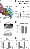
Biogenesis of mitochondrial iron-sulfur (Fe/S) cluster proteins requires the interaction of multiple proteins with the highly conserved 14-kDa scaffold protein Isu, on which clusters are built prior to their transfer to recipient proteins. For example, the assembly process requires the cysteine desulfurase Nfs1, which serves as the sulfur donor for cluster assembly. The transfer process requires Jac1, a J-protein Hsp70 cochaperone. We recently identified three residues on the surface of Jac1 that form a hydrophobic patch critical for interaction with Isu. The results of molecular modeling of the Isu1-Jac1 interaction, which was guided by these experimental data and structural/biophysical information available for bacterial homologs, predicted the importance of three hydrophobic residues forming a patch on the surface of Isu1 for interaction with Jac1. Using Isu variants having alterations in residues that form the hydrophobic patch on the surface of Isu, this prediction was experimentally validated by in vitro binding assays. In addition, Nfs1 was found to require the same hydrophobic residues of Isu for binding, as does Jac1, suggesting that Jac1 and Nfs1 binding is mutually exclusive. In support of this conclusion, Jac1 and Nfs1 compete for binding to Isu. Evolutionary analysis revealed that residues involved in these interactions are conserved and that they are critical residues for the biogenesis of Fe/S cluster protein in vivo. We propose that competition between Jac1 and Nfs1 for Isu binding plays an important role in transitioning the Fe/S cluster biogenesis machinery from the cluster assembly step to the Hsp70-mediated transfer of the Fe/S cluster to recipient proteins.
Keywords: ISC System; Iron-Sulfur Protein; J-protein; Mitochondria; Molecular Chaperone; Scaffold Proteins; Yeast.
Figures


Similar articles
-
Overlapping binding sites of the frataxin homologue assembly factor and the heat shock protein 70 transfer factor on the Isu iron-sulfur cluster scaffold protein.J Biol Chem. 2014 Oct 31;289(44):30268-30278. doi: 10.1074/jbc.M114.596726. Epub 2014 Sep 16. J Biol Chem. 2014. PMID: 25228696 Free PMC article.
-
Interaction of J-protein co-chaperone Jac1 with Fe-S scaffold Isu is indispensable in vivo and conserved in evolution.J Mol Biol. 2012 Mar 16;417(1-2):1-12. doi: 10.1016/j.jmb.2012.01.022. Epub 2012 Jan 27. J Mol Biol. 2012. PMID: 22306468 Free PMC article.
-
Frataxin directly stimulates mitochondrial cysteine desulfurase by exposing substrate-binding sites, and a mutant Fe-S cluster scaffold protein with frataxin-bypassing ability acts similarly.J Biol Chem. 2013 Dec 27;288(52):36773-86. doi: 10.1074/jbc.M113.525857. Epub 2013 Nov 11. J Biol Chem. 2013. PMID: 24217246 Free PMC article.
-
Molecular chaperones HscA/Ssq1 and HscB/Jac1 and their roles in iron-sulfur protein maturation.Crit Rev Biochem Mol Biol. 2007 Mar-Apr;42(2):95-111. doi: 10.1080/10409230701322298. Crit Rev Biochem Mol Biol. 2007. PMID: 17453917 Review.
-
Mechanisms of Mitochondrial Iron-Sulfur Protein Biogenesis.Annu Rev Biochem. 2020 Jun 20;89:471-499. doi: 10.1146/annurev-biochem-013118-111540. Epub 2020 Jan 14. Annu Rev Biochem. 2020. PMID: 31935115 Review.
Cited by
-
Role of Nfu1 and Bol3 in iron-sulfur cluster transfer to mitochondrial clients.Elife. 2016 Aug 17;5:e15991. doi: 10.7554/eLife.15991. Elife. 2016. PMID: 27532773 Free PMC article.
-
PANoptosis in cancer, the triangle of cell death.Cancer Med. 2023 Dec;12(24):22206-22223. doi: 10.1002/cam4.6803. Epub 2023 Dec 8. Cancer Med. 2023. PMID: 38069556 Free PMC article. Review.
-
Architecture of the Yeast Mitochondrial Iron-Sulfur Cluster Assembly Machinery: THE SUB-COMPLEX FORMED BY THE IRON DONOR, Yfh1 PROTEIN, AND THE SCAFFOLD, Isu1 PROTEIN.J Biol Chem. 2016 May 6;291(19):10378-98. doi: 10.1074/jbc.M115.712414. Epub 2016 Mar 3. J Biol Chem. 2016. PMID: 26941001 Free PMC article.
-
Evolutionary conservation and in vitro reconstitution of microsporidian iron-sulfur cluster biosynthesis.Nat Commun. 2017 Jan 4;8:13932. doi: 10.1038/ncomms13932. Nat Commun. 2017. PMID: 28051091 Free PMC article.
-
Architecture of the Human Mitochondrial Iron-Sulfur Cluster Assembly Machinery.J Biol Chem. 2016 Sep 30;291(40):21296-21321. doi: 10.1074/jbc.M116.738542. Epub 2016 Aug 12. J Biol Chem. 2016. PMID: 27519411 Free PMC article.
References
-
- Lill R., Hoffmann B., Molik S., Pierik A. J., Rietzschel N., Stehling O., Uzarska M. A., Webert H., Wilbrecht C., Mühlenhoff U. (2012) The role of mitochondria in cellular iron-sulfur protein biogenesis and iron metabolism. Biochim. Biophys. Acta 1823, 1491–1508 - PubMed
-
- Garland S. A., Hoff K., Vickery L. E., Culotta V. C. (1999) Saccharomyces cerevisiae ISU1 and ISU2. Members of a well-conserved gene family for iron-sulfur cluster assembly. J. Mol. Biol. 294, 897–907 - PubMed
-
- Tsai C. L., Barondeau D. P. (2010) Human frataxin is an allosteric switch that activates the Fe-S cluster biosynthetic complex. Biochemistry 49, 9132–9139 - PubMed
Publication types
MeSH terms
Substances
Grants and funding
LinkOut - more resources
Full Text Sources
Other Literature Sources
Molecular Biology Databases
Research Materials
Miscellaneous

