

Cytochrome p450 cyp26a1 alters spinal motor neuron subtype identity in differentiating embryonic stem cells
- PMID: 23946489
- PMCID: PMC3789976
- DOI: 10.1074/jbc.M113.474254
Cytochrome p450 cyp26a1 alters spinal motor neuron subtype identity in differentiating embryonic stem cells
Abstract
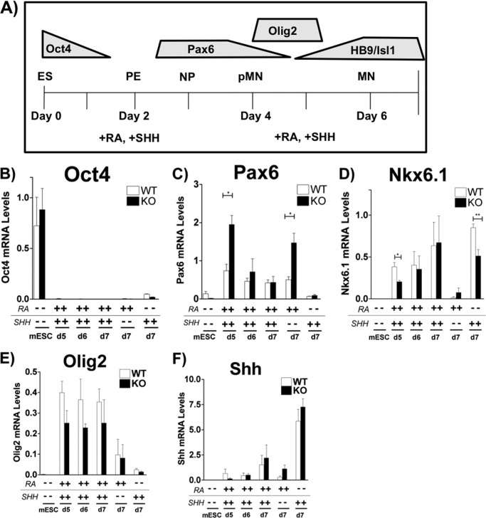
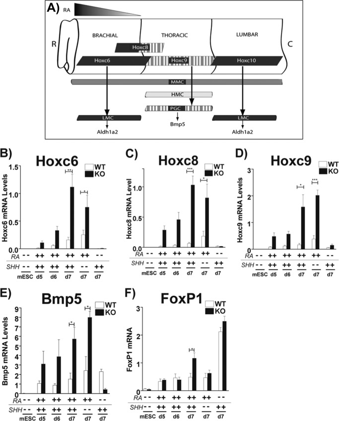
The ability to differentiate embryonic stem cells (ESCs) into specific cell types is critical for improved regenerative medicine strategies, cancer chemotherapeutic approaches, and regimens to combat chronic diseases associated with aging. Subclasses of motor neurons (MNs) are generated at different positions along the rostrocaudal axis of the spinal cord, and the signals that specify MN subtype fates remain poorly defined. We show here that the cytochrome P450 enzyme Cyp26a1, which metabolizes all-trans-retinoic acid (RA) and thereby reduces RA levels, plays a crucial role in specifying MN columnar subtypes. Lack of Cyp26a1 in ESCs during differentiation to spinal MNs increases Aldh1a2 (RALDH2) and Hoxc6, markers of the Hox-dependent, lateral motor column (LMC) subtype identity. In contrast, Lhx3, a marker for median motor column identity, showed lower expression in Cyp26a1(-/-)-derived MNs compared with WT. Without Cyp26a1, an increase in intracellular RA concentration plus sonic hedgehog agonist treatment confer an LMC fate on differentiating MNs. Our data suggest a strategy for increasing LMC-type MNs from ESCs by blocking Cyp26a1 in cell replacement/ESC differentiation therapy to treat neurodegenerative diseases, such as amyotrophic lateral sclerosis.
Keywords: Cytochrome P450; Differentiation; Neurological Diseases; Retinoid; Stem Cells.
Figures


Similar articles
-
CYP26A1 knockout embryonic stem cells exhibit reduced differentiation and growth arrest in response to retinoic acid.Dev Biol. 2008 Mar 15;315(2):331-54. doi: 10.1016/j.ydbio.2007.12.021. Epub 2007 Dec 27. Dev Biol. 2008. PMID: 18241852
-
Retinoic Acid-Mediated Regulation of GLI3 Enables Efficient Motoneuron Derivation from Human ESCs in the Absence of Extrinsic SHH Activation.J Neurosci. 2015 Aug 19;35(33):11462-81. doi: 10.1523/JNEUROSCI.3046-14.2015. J Neurosci. 2015. PMID: 26290227 Free PMC article.
-
Ethanol promotes differentiation of embryonic stem cells through retinoic acid receptor-γ.J Biol Chem. 2019 Apr 5;294(14):5536-5548. doi: 10.1074/jbc.RA118.007153. Epub 2019 Feb 8. J Biol Chem. 2019. PMID: 30737277 Free PMC article.
-
Cytochrome P450s in the regulation of cellular retinoic acid metabolism.Annu Rev Nutr. 2011 Aug 21;31:65-87. doi: 10.1146/annurev-nutr-072610-145127. Annu Rev Nutr. 2011. PMID: 21529158 Free PMC article. Review.
-
Establishing and maintaining Hox profiles during spinal cord development.Semin Cell Dev Biol. 2024 Jan-Feb;152-153:44-57. doi: 10.1016/j.semcdb.2023.03.014. Epub 2023 Apr 5. Semin Cell Dev Biol. 2024. PMID: 37029058 Free PMC article. Review.
Cited by
-
Cytochrome P450 26A1 Contributes to the Maintenance of Neuropathic Pain.Neurosci Bull. 2024 Mar;40(3):293-309. doi: 10.1007/s12264-023-01101-1. Epub 2023 Aug 28. Neurosci Bull. 2024. PMID: 37639183 Free PMC article.
-
Retinoic acid suppresses the canonical Wnt signaling pathway in embryonic stem cells and activates the noncanonical Wnt signaling pathway.Stem Cells. 2014 Aug;32(8):2061-71. doi: 10.1002/stem.1706. Stem Cells. 2014. PMID: 24648413 Free PMC article.
-
Combinatorial knockout of RARα, RARβ, and RARγ completely abrogates transcriptional responses to retinoic acid in murine embryonic stem cells.J Biol Chem. 2018 Jul 27;293(30):11891-11900. doi: 10.1074/jbc.RA118.001951. Epub 2018 May 30. J Biol Chem. 2018. PMID: 29848550 Free PMC article.
-
Human cytochrome P450 enzymes 5-51 as targets of drugs and natural and environmental compounds: mechanisms, induction, and inhibition - toxic effects and benefits.Drug Metab Rev. 2018 Aug;50(3):256-342. doi: 10.1080/03602532.2018.1483401. Drug Metab Rev. 2018. PMID: 30717606 Free PMC article. Review.
-
Retinoid metabolism: new insights.J Mol Endocrinol. 2022 Oct 11;69(4):T37-T49. doi: 10.1530/JME-22-0082. Print 2022 Nov 1. J Mol Endocrinol. 2022. PMID: 35900851 Free PMC article. Review.
References
-
- Ludolph A. C., Brettschneider J., Weishaupt J. H. (2012) Amyotrophic lateral sclerosis. Curr. Opin. Neurol. 25, 530–535 - PubMed
-
- Pasinelli P., Brown R. H. (2006) Molecular biology of amyotrophic lateral sclerosis. Insights from genetics. Nat. Rev. Neurosci. 7, 710–723 - PubMed
-
- Bruijn L. I., Miller T. M., Cleveland D. W. (2004) Unraveling the mechanisms involved in motor neuron degeneration in ALS. Annu. Rev. Neurosci. 27, 723–749 - PubMed
-
- López-González R., Kunckles P., Velasco I. (2009) Transient recovery in a rat model of familial amyotrophic lateral sclerosis after transplantation of motor neurons derived from mouse embryonic stem cells. Cell Transplant. 18, 1171–1181 - PubMed
-
- Dimos J. T., Rodolfa K. T., Niakan K. K., Weisenthal L. M., Mitsumoto H., Chung W., Croft G. F., Saphier G., Leibel R., Goland R., Wichterle H., Henderson C. E., Eggan K. (2008) Induced pluripotent stem cells generated from patients with ALS can be differentiated into motor neurons. Science 321, 1218–1221 - PubMed
Publication types
MeSH terms
Substances
Grants and funding
LinkOut - more resources
Full Text Sources
Other Literature Sources
