

Case Reports
doi: 10.1136/bcr-2013-010438.
Spindle cell lipoma
Affiliations
- PMID: 23946512
- PMCID: PMC3762247
- DOI: 10.1136/bcr-2013-010438
Item in Clipboard
Case Reports
Spindle cell lipoma
BMJ Case Rep.
.
Abstract
Spindle cell lipomas (SCLs) are a group of benign lipogenic tumours, typically arising in the posterior neck, upper back and shoulder of elderly male patients. Approximately 80% of these tumours arise in characteristic location, but 20% arise in unusual locations, thereby making these cases more difficult to diagnose. We present a case of SCL occurring in the right periorbital region of a 14-year-old boy. The MRI was suggestive of possible malignancy. Diagnosis of neurofibroma was made on incisional biopsy. However, the histopathological and immunohistochemical analyses of the excised lesion confirmed the diagnosis of SCL.
Figures

Photograph showing swelling around the right eye causing compression of the eye.

Photograph showing swelling and increased hair growth over the skin of involved area.

Coronal CT image showing soft tissue density lesion along the lateral wall of right orbit involving temporalis muscle.

Coronal and axial MRI T2-weighted images showing heterogeneous area of altered signal intensity involving the temporalis muscle.

Intraoperative photograph.
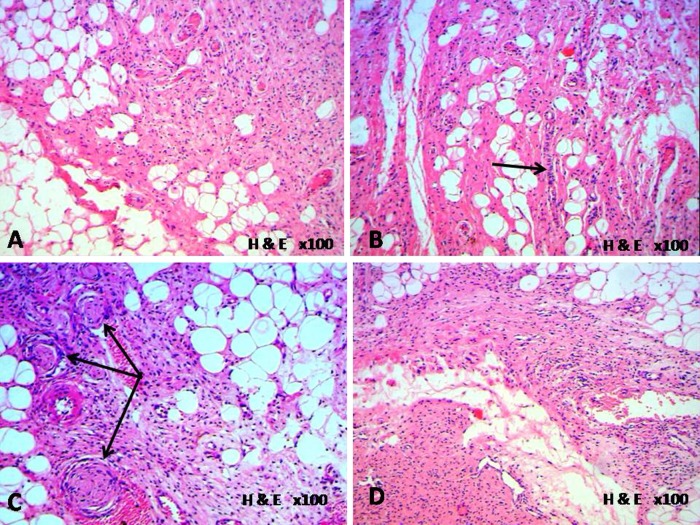
Photomicrograph showing histopathological features. (A) predominance of spindle component; (B) parallel arrangement of spindle cells; (C) whorled pattern of spindle cells and (D) myxoid area.

High power views showing bland and uniform short spindle cells along with mast cells in a fibrous stroma.

Immunohistochemistry photomicrographs showing strong and diffused positivity of spindle cells for CD34.

Immunohistochemistry photomicrograph showing negative staining of spindle cells for S-100 protein.
References
-
- Enzinger FM, Harvey DA. Spindle-cell lipoma. Cancer 1975;2013:1852–9 - PubMed
-
- Fletcher CDM, Martin-Bates E. Spindle-cell lipoma: a clinicopathological study with some original observations. Histopathology 1987;2013:803–17 - PubMed
-
- Comunoglu N, Comunoglu C, Ekic AI, et al. Spindle cell lipoma. Pol J Pathol 2007;2013:7–11 - PubMed
-
- Weiss SW, Goldblum JR, eds. Benign lipomatous tumors. In: Enzinger and Weiss's soft tissue tumors, 4th edn. St. Louis: Mosby, 2001;571–639
-
- Syed S, Martin AM, Haupt H, et al. Frequent detection of androgen receptors in spindle cell lipomas: an explanation for this lesion's male predominance? Arch Pathol Lab Med 2008;2013:81–3 - PubMed
Publication types
MeSH terms
LinkOut - more resources
Full Text Sources
Other Literature Sources
Research Materials
Miscellaneous