

Cell-specific transcriptional profiling reveals candidate mechanisms regulating development and function of uterine epithelia in mice
- PMID: 23946541
- PMCID: PMC7289334
- DOI: 10.1095/biolreprod.113.111971
Cell-specific transcriptional profiling reveals candidate mechanisms regulating development and function of uterine epithelia in mice
Abstract
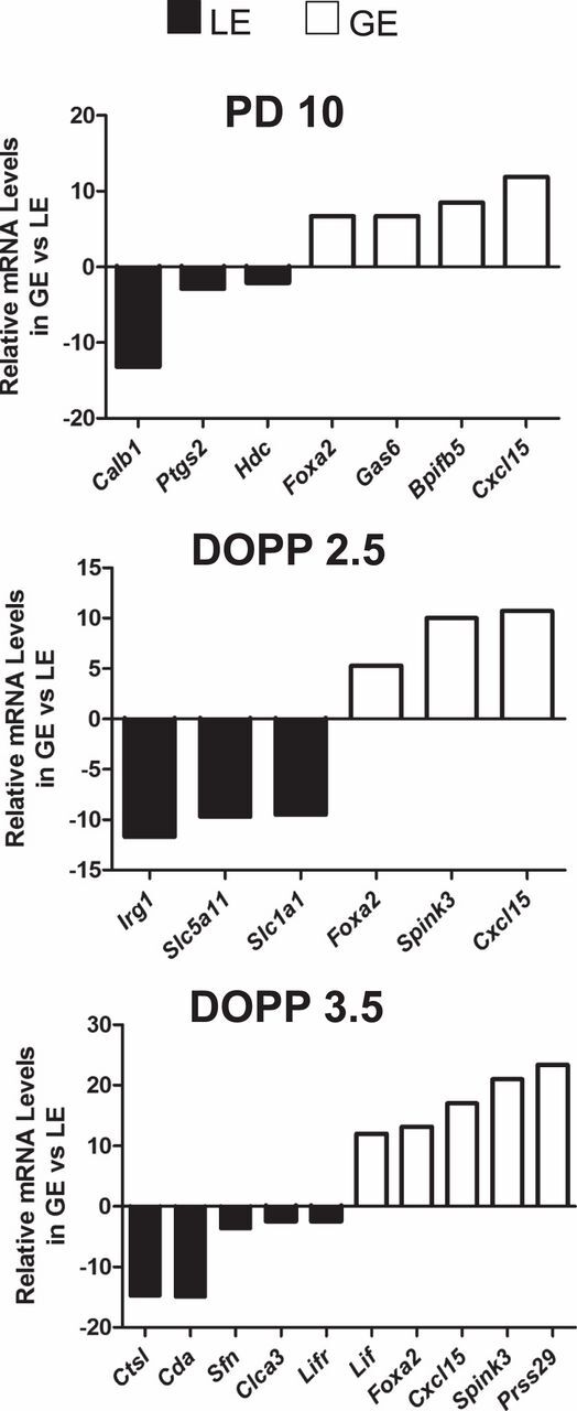
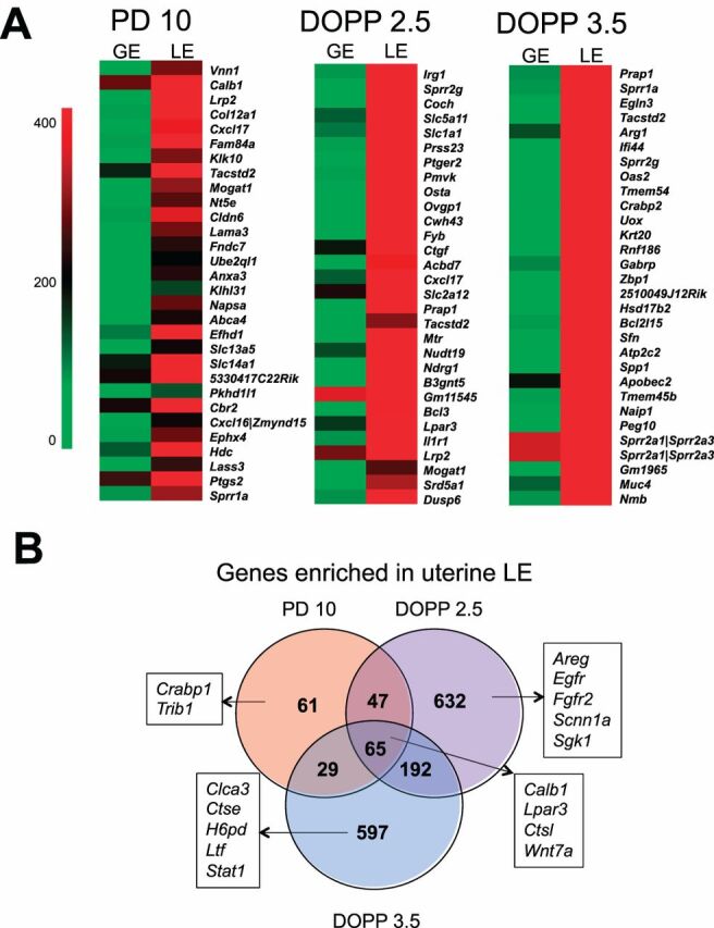
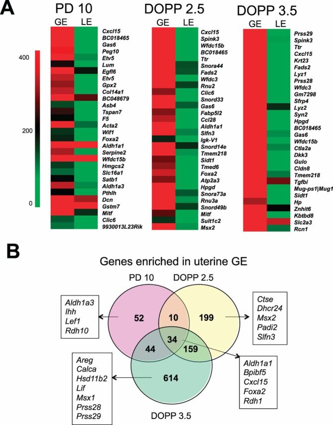
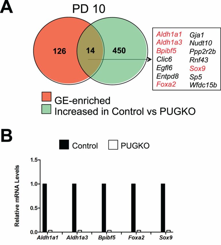
All mammalian uteri have luminal (LE) and glandular epithelia (GE) in their endometrium. The LE mediates uterine receptivity and blastocyst attachment for implantation, and the GE synthesize and secrete or transport bioactive substances involved in blastocyst implantation, uterine receptivity, and stromal cell decidualization. However, the mechanisms governing uterine epithelial development after birth and their function in the adult are not fully understood. Here, comprehensive microarray analysis was conducted on LE and GE isolated by laser capture microdissection from uteri on Postnatal Day 10 (PD 10) and day of pseudopregnancy (DOPP) 2.5 and 3.5. This data was integrated with analysis of uteri from gland-containing control and aglandular progesterone-induced uterine gland knockout mice from PD 10 and DOPP 3.5. Many genes were expressed in both epithelia, but there was greater expression of genes in the LE than in the GE. In the neonate, GE-expressed genes were enriched for morphogenesis, development, migration, and retinoic acid signaling. In the adult, LE-expressed genes were enriched for metabolic processes and steroid biosynthesis, whereas retinoid signaling, tight junction, extracellular matrix, and regulation of kinase activity were enriched in the GE. The transcriptome differences in the epithelia support the idea that each cell type has a distinct and complementary function in the uterus. The candidate genes and regulatory networks identified here provide a framework to discover new mechanisms regulating development of epithelia in the postnatal uterus and their functions in early pregnancy.
Keywords: endometrium; gene expression; guinea pigs; implantation; mice; rodents (rats; uterus; voles).
Figures
References
-
- Brody JR,Cunha GR.. Histologic, morphometric, and immunocytochemical analysis of myometrial development in rats and mice: I. Normal development.Am J Anat 1989;186:1–20. - PubMed
-
- Bartol FF,Wiley AA,Floyd JG,Ott TL,Bazer FW,Gray CA,Spencer TE.. Uterine differentiation as a foundation for subsequent fertility.J Reprod Fertil Suppl 1999;54:287–302. - PubMed
-
- Hu J,Gray CA,Spencer TE.. Gene expression profiling of neonatal mouse uterine development.Biol Reprod 2004;70:1870–1876. - PubMed
-
- Branham WS,Sheehan DM,Zehr DR,Ridlon E,Nelson CJ.. The postnatal ontogeny of rat uterine glands and age-related effects of 17 beta-estradiol.Endocrinology 1985;117:2229–2237. - PubMed
-
- Spencer TE,Dunlap KA,Filant J.. Comparative developmental biology of the uterus: insights into mechanisms and developmental disruption.Mol Cell Endocrinol 2012;354:34–53. - PubMed
Publication types
MeSH terms
Substances
Grants and funding
LinkOut - more resources
Full Text Sources
Other Literature Sources
Medical
Molecular Biology Databases
