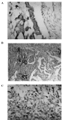
Correlation of the expression of vascular endothelial growth factor and its receptors with microvessel density in ovarian cancer
- PMID: 23946799
- PMCID: PMC3742816
- DOI: 10.3892/ol.2013.1349
Correlation of the expression of vascular endothelial growth factor and its receptors with microvessel density in ovarian cancer
Abstract
The present study aimed to investigate the correlation between the expression of vascular endothelial growth factor (VEGF) and its receptors, the Flt-1 and KDR proteins, with clinical pathology and microvessel density (MVD) in ovarian cancer tissue. The protein expression levels of VEGF and its receptors, Flt-1 and KDR/Flk-1, were detected in 48 cases of ovarian cancer using the streptavidin-biotin complex (SABC) immunohistochemical method, and tumor MVD was evaluated using F8 factor (FVIII-RA). The expression of the VEGF, Flt-1 and KDR proteins was not significantly associated with the pathological type, extent of differentiation or clinical stage of ovarian cancer. However, the co-expression of VEGF and Flt-1 was markedly correlated with differentiation and clinical stage (P<0.01). The co-expression levels of VEGF and receptor Flt-1 in malignant neoplasms with lymph node metastasis was significantly higher compared with malignant neoplasms without lymph node metastasis (P<0.05). The expression level of KDR in patients with hepatic metastasis was significantly higher compared with patients without hepatic metastasis (P<0.05). The co-expression level of VEGF and KDR in patients with hepatic metastasis was significantly higher compared with patients without hepatic metastasis (P<0.05) and the Flt-1 expression level in patients with ascites <1,000 ml was significantly lower than that in patients with ascites ≥1000 ml (P<0.05). The mean MVD of VEGF- and KDR-positive patients was significantly higher compared with VEGF- and KDR-negative patients (P<0.05). The expression of VEGF and its receptors is involved in the malignant transformation of ovarian tumors, tumor progression and metastasis, as well as ascites formation and angiogenesis.
Keywords: Flt-1 receptor; KDR receptor; ascites; oophoroma; vascular endothelial growth factor.
Figures


Similar articles
-
Expression of vascular endothelial growth factor and its receptors flt and KDR in ovarian carcinoma.J Natl Cancer Inst. 1995 Apr 5;87(7):506-16. doi: 10.1093/jnci/87.7.506. J Natl Cancer Inst. 1995. PMID: 7707437
-
Immunolocalizations of VEGF, its receptors flt-1, KDR and TGF-beta's in epithelial ovarian tumors.Histol Histopathol. 2006 Oct;21(10):1055-64. doi: 10.14670/HH-21.1055. Histol Histopathol. 2006. PMID: 16835828
-
VEGF, flt-1, and KDR/flk-1 as prognostic indicators in endometrial carcinoma.Gynecol Oncol. 2000 Jan;76(1):33-9. doi: 10.1006/gyno.1999.5658. Gynecol Oncol. 2000. PMID: 10620438
-
Study of microvessel density and the expression of the angiogenic factors VEGF, bFGF and the receptors Flt-1 and FLK-1 in benign, premalignant and malignant prostate tissues.Histol Histopathol. 2006 Aug;21(8):857-65. doi: 10.14670/HH-21.857. Histol Histopathol. 2006. PMID: 16691538
-
The vascular endothelial growth factor receptor KDR/Flk-1 is a major regulator of malignant ascites formation in the mouse hepatocellular carcinoma model.Hepatology. 2001 Apr;33(4):841-7. doi: 10.1053/jhep.2001.23312. Hepatology. 2001. PMID: 11283848
Cited by
-
Vascular invasion in hepatitis B virus-related hepatocellular carcinoma with underlying cirrhosis: possible associations with ascites and hepatitis B viral factors?Tumour Biol. 2015 Aug;36(8):6255-63. doi: 10.1007/s13277-015-3311-8. Epub 2015 Apr 2. Tumour Biol. 2015. PMID: 25833692
-
PARP-1 may be involved in angiogenesis in epithelial ovarian cancer.Oncol Lett. 2016 Dec;12(6):4561-4567. doi: 10.3892/ol.2016.5226. Epub 2016 Oct 5. Oncol Lett. 2016. PMID: 28101214 Free PMC article.
-
Expression and clinical significance of vascular endothelial growth factor and fms-related tyrosine kinase 1 in colorectal cancer.Oncol Lett. 2015 May;9(5):2414-2418. doi: 10.3892/ol.2015.3013. Epub 2015 Mar 4. Oncol Lett. 2015. PMID: 26137082 Free PMC article.
-
MicroRNA-126 affects ovarian cancer cell differentiation and invasion by modulating expression of vascular endothelial growth factor.Oncol Lett. 2018 Apr;15(4):5803-5808. doi: 10.3892/ol.2018.8025. Epub 2018 Feb 12. Oncol Lett. 2018. PMID: 29552211 Free PMC article.
-
Factors influencing improvement of visual field after trans-sphenoidal resection of pituitary macroadenomas: a retrospective cohort study.Int J Ophthalmol. 2015 Dec 18;8(6):1224-8. doi: 10.3980/j.issn.2222-3959.2015.06.27. eCollection 2015. Int J Ophthalmol. 2015. PMID: 26682178 Free PMC article.
References
-
- Nsihida N, Yano H, Komai K, Nishida T, Kamura T, Kojiro M. Vascular endothelial growth factor-C and vascular endothelial growth factor receptor 2 are related closely to the prognosis of patients with ovarian carcinoma. Cancer. 2004;101:1364–1374. - PubMed
-
- Karavasilis V, Malamou-Mitsi V, Briasoulis E, Tsanou E, Kitsou E, Pavlidis N. Clinicopathologic study of vascular endothelial growth factor, thrombospondin-1, and microvessel density assessed by CD34 in patients with stage III ovarian carcinoma. Int J Gynecol Cancer. 2006;16(Suppl 1):241–246. - PubMed
-
- Gómez-Raposo C, Mendiola M, Barriuso J, Casado E, Hardisson D, Redondo A. Angiogenesis and ovarian cancer. Clin Transl Oncol. 2009;11:564–571. - PubMed
-
- Diniz Bizzo SM, Meira DD, Lima JM, Mororó Jda S, Casali-da-Rocha JC, Ornellas MH. Peritoneal VEGF burden as a predictor of cytoreductive surgery outcome in women with epithelial ovarian cancer. Int J Gynaecol Obstet. 2010;109:113–117. - PubMed
-
- Hefler LA, Zeillinger R, Grimm C, et al. Preoperative serum vascular endothelial growth factor as a prognostic parameter in ovarian cancer. Gynecol Oncol. 2006;103:512–517. - PubMed
LinkOut - more resources
Full Text Sources
Other Literature Sources
Miscellaneous
