Review
. 2013 Aug 27;52(34):5708-22.
doi: 10.1021/bi400804z. Epub 2013 Aug 15.

MscS-like mechanosensitive channels in plants and microbes

Affiliations
Review

MscS-like mechanosensitive channels in plants and microbes

Margaret E Wilson et al. Biochemistry. .

Abstract

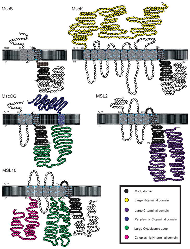
The challenge of osmotic stress is something all living organisms must face as a result of environmental dynamics. Over the past three decades, innovative research and cooperation across disciplines have irrefutably established that cells utilize mechanically gated ion channels to release osmolytes and prevent cell lysis during hypoosmotic stress. Early electrophysiological analysis of the inner membrane of Escherichia coli identified the presence of three distinct mechanosensitive activities. The subsequent discoveries of the genes responsible for two of these activities, the mechanosensitive channels of large (MscL) and small (MscS) conductance, led to the identification of two diverse families of mechanosensitive channels. The latter of these two families, the MscS family, consists of members from bacteria, archaea, fungi, and plants. Genetic and electrophysiological analysis of these family members has provided insight into how organisms use mechanosensitive channels for osmotic regulation in response to changing environmental and developmental circumstances. Furthermore, determining the crystal structure of E. coli MscS and several homologues in several conformational states has contributed to our understanding of the gating mechanisms of these channels. Here we summarize our current knowledge of MscS homologues from all three domains of life and address their structure, proposed physiological functions, electrophysiological behaviors, and topological diversity.


Figures

Figure 1
Figure 1. Schematic representation of models for mechanosensitive channel gating
(A) The intrinsic bilayer model, wherein lateral membrane tension favors the open state of the channel. (B) The tethered trapdoor model, wherein a tether to an extracellular (in this case) component exerts force on the channel, leading to its gating. (C) The unified model, wherein a tether to an extracellular component leads to reorientation of the channel within the membrane bilayer, thereby gating it.
Figure 2
Figure 2. Crystal structures of E. coli MscS and homologs
(A) EcMscS in inactive/non-conductive state (2OAU, Steinbacher, 2007); (B) TtMscS from T. tengcongensis in closed state (3UDC, Zhang, 2012); (C) A106V EcMscS mutant in open state (2VV5, Wang, 2008); (D) EcMscS in open state (4HWA, Lai, 2013). Left panel, side view of the heptameric channel. Middle panel, view from the periplasmic side. Right panel, space-filling representation of the pore from the periplasmic side; the channels are truncated at I175 (for EcMscS) for an unobstructed view. Basic residues are blue, acidic residues are red, polar residues are green, non-polar residues are white. The images were generated with VMD software (University of Illinois).
Figure 3
Figure 3. The conserved region of EcMscS and TtMscS monomers in different conformations
A single monomer of (A) EcMscS (aa 27-175) in a nonconducting state (2OAU, Steinbacher, 2007); (B) TtMscS (aa 13-175) in a nonconducting state (3UDC, Zhang, 2012); (C) EcMscS A106V (aa 25-175) in an open state (2VV5, Wang, 2008). (D) Superposition of panel A with a single monomer of EcMscS (27-175) in an open state (4HWA, Lai, 2013). The kink-forming residues G113 (EcMscS) and G109 (TtMscS) are represented as blue spheres and the A106V mutation as a red sphere. The vapor-lock residues L105 and L109 are labeled in yellow. F68 and L111, residues proposed to mediate the TM2-TM3 interaction (Belyy, 2010) are labeled magenta. Images were generated with VMD software (University of Illinois).
Figure 4
Figure 4. Monomer topologies of representative MscS family members
MscS monomer topology was rendered based on Naismith and Booth, 2012. The domain conserved among all MscS homologs is indicated in black; other domains are colored as indicated in the legend. For the purpose of clarity TM3b of MscS is represented outside the lipid bilayer. MscK and MscCG topologies were predicted with TOPCONS (http://topcons.net/) and ARAMEMNON (http://aramemnon.botanik.uni-koeln.de/) for MSL2 and MSL10. Processed versions of MscK and MSL2 are presented.

Similar articles

Cited by

References

    1. Gadsby DC. Ion channels versus ion pumps: the principal difference, in principle. Nat Rev Mol Cell Biol. 2009;10:344–352. - PMC - PubMed
    1. Hille B. Ion channels of excitable membranes. 3. Sinauer; Sunderland, Mass: 2001.
    1. Ringer S. Regarding the Action of Hydrate of Soda, Hydrate of Ammonia, and Hydrate of Potash on the Ventricle of the Frog’s Heart. J Physiol. 1882;3:195–202. 196. - PMC - PubMed
    1. Takahashi K, Kakimoto Y, Toda K, Naruse K. Mechanobiology in cardiac physiology and diseases. J Cell Mol Med. 2013;17:225–232. - PMC - PubMed
    1. Kullmann DM. Neurological channelopathies. Annu Rev Neurosci. 2010;33:151–172. - PubMed

Publication types

MeSH terms

LinkOut - more resources