. 2013 Sep 17;52(37):6456-66.
doi: 10.1021/bi400682n. Epub 2013 Aug 30.

Interaction of profilin with the barbed end of actin filaments

Affiliations

Interaction of profilin with the barbed end of actin filaments

Naomi Courtemanche et al. Biochemistry. .

Abstract

Profilin binds not only to actin monomers but also to the barbed end of the actin filament, where it inhibits association of subunits. To address open questions about the interactions of profilin with barbed ends, we measured the effects of a wide range of concentrations of Homo sapiens profilin 1 on the rate of elongation of individual skeletal muscle actin filaments by total internal reflection fluorescence microscopy. Much higher concentrations of profilin were required to stop elongation by AMP-PNP-actin monomers than ADP-actin monomers. High concentrations of profilin depolymerized barbed ends at a rate much faster than the spontaneous dissociation rates of Mg-ATP-, Mg-AMP-PNP-, Mg-ADP-Pi-, and Mg-ADP-actin subunits. Fitting a thermodynamic model to these data allowed us to determine the affinities of profilin and profilin-actin for barbed ends and the influence of the nucleotide bound to actin on these interactions. Profilin has a much higher affinity for ADP-actin filament barbed ends (Kd = 1 μM) than AMP-PNP-actin filament barbed ends (Kd = 226 μM). ADP-actin monomers associated with profilin bind to ADP-actin filament barbed ends 10% as fast as free ADP-actin monomers, but bound profilin does not affect the rate of association of AMP-PNP-actin monomers with barbed ends. The differences in the affinities of AMP-PNP- and ADP-bound barbed ends for profilin and profilin-actin suggest that conformations of barbed end subunits differ from those of monomers and change upon nucleotide hydrolysis and phosphate release. A structural model revealed minor steric clashes between profilin and actin subunits at the barbed end that explain the biochemical results.


Figures

Figure 1
Figure 1
Thermodynamic scheme of actin filament elongation in the presence of profilin. Four reactions are depicted and labeled as follows: (1) reaction of profilin (P) with an actin monomer (A); (2) reaction of an actin monomer with the barbed ends of a filament (BE); (3) reaction of profilin with the barbed end of a filament; and (4) reaction of the profilin-actin complex (PA) with the barbed end of a filament. “F” represents the newly incorporated filamentous subunit upon actin or profilin-actin binding to the barbed end. “BE-P” represents the profilin-bound barbed end. To satisfy a detailed balance, the sum of the free energies of reactions 1 and 4 must be equal to the sum of the free energies of reactions 2 and 3. Table 2 lists the thermodynamic and kinetic parameters for AMP-PNP-actin, ADP-Pi-actin and ADP-actin.
Figure 2
Figure 2
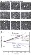
Effect of profilin on barbed end elongation of Mg-AMP-PNP-actin filaments. Polymerization conditions: 10 mM imidazole (pH 7.0), 50 mM KCl, 1 mM MgCl2, 1 mM EGTA, 50 mM DTT, 0.37 mM AMP-PNP, 0.02 mM CaCl2, 15 mM glucose, 0.02 mg/ml catalase, 0.1 mg/ml glucose oxidase and 0.5% methylcellulose (4,000 cP at 2% (w/v)). Data were collected with total internal reflection fluorescence microscopy. (A) Time-series of images of 1.5 μM AMP-PNP-actin (20% Alexa 488-labeled) filaments growing in the presence of a range of concentrations of human profilin 1. Yellow arrowheads denote barbed ends. (B) Time courses of the growth of six representative filament barbed ends in the presence of 1.5 μM AMP-PNP-actin (20% Alexa 488-labeled) and profilin concentrations of 0 (black lines), 10 μM (red lines) or 225 μM (blue lines).
Figure 3
Figure 3
Dependence of the elongation rates of AMP-PNP-actin filaments on the concentration of AMP-PNP-actin monomers. Growth of filaments was observed by total internal reflection fluorescence microscopy with concentrations of AMP-PNP-actin (20% Alexa-488-labeled) from 50 nM to 2.5 μM and in microscopy buffer with 0.37 mM AMP-PNP. Filament seeds were grown from 1.5 μM AMP-PNP-actin monomers in the observation chamber, followed by a wash and replacement with AMP-PNP-actin monomers. Error bars are 1 standard error of the mean elongation rate of at least 10 filaments.
Figure 4
Figure 4
Dependence of barbed end elongation rates by Mg-ATP-, Mg-AMP-PNP-, Mg-ADP-Pi- and Mg-ADP-actin monomers on the concentration of profilin 1. Data were collected by total internal reflection fluorescence microscopy. Error bars are standard errors of the mean elongation rates of at least 10 filaments. The smooth curves are fits to the filled data points with the model described in the text. The open data points were masked in these fits. (A) Conditions for Mg-ATP-actin monomers: 1.5 μM ATP-actin (20% Alexa-488-labeled), 0.2 mM ATP and a range of concentrations of profilin in microscopy buffer. (B) Conditions for Mg-AMP-PNP-actin monomers: 1.5 μM AMP-PNP-actin (20% Alexa-488-labeled), 0.37 mM AMP-PNP and a range of concentrations of profilin in microscopy buffer. (C) Conditions for Mg-ADP-Pi-actin monomers: 4 μM Mg-ADP-Pi-actin (20% Alexa-488-labeled), 0.3 mM ADP, 20 units/mL hexokinase, 12.5 mM potassium phosphate and a range of concentrations of profilin in microscopy buffer. The polymerization buffer contained 25 mM KCl instead of 50 mM to compensate for the additional K+ ions. (D) Conditions for Mg-ADP-actin monomers: 5.0 μM Mg-ADP-actin (20% Alexa-488-actin), 0.3 mM ADP and 20 units/mL hexokinase and a range of concentrations of profilin in microscopy buffer.
Figure 5
Figure 5
Molecular models showing steric clashes produced by profilin binding to the terminal subunits of both long-pitch helices (subunits n and n−1) of an actin filament. Solvent excluded surface model of actin and ribbon diagrams of profilin are shown. (A) Superposition of human profilin (purple; pdb: 1FIK) onto the structure of bovine β-actin (green) bound with bovine profilin (blue; pdb: 1HLU). (B) (Upper panels) Stereo view of an Oda actin filament model (30) with human profilin I (dark purple and light purple) bound to the terminal subunits of each long-pitch helix (subunits n and n−1) as in (A). (Lower panels) Additional views of the filament and bound profilin molecules. (C) Detail of the clashes formed between profilin (dark purple) and subdomain 1 of actin subunit n (yellow) when profilin is bound to actin subunit n−1. Solvent excluded surfaces are shown (33). (D) Detail of the profilin-actin interface at the terminal barbed end subunit (profilin, light purple; actin subunit n, yellow). Solvent excluded surfaces are shown (33).

References

    1. Birbach A. Profilin, a multi-modal regulator of neuronal plasticity. Bioessays. 2008;30:994–1002. - PubMed
    1. Carlsson L, Nyström LE, Sundkvist I, Markey F, Lindberg U. Actin polymerizability is influenced by profilin, a low molecular weight protein in non-muscle cells. J Mol Biol. 1977;115:465–483. - PubMed
    1. Markey F, Larsson H, Weber K, Lindberg U. Nucleation of actin polymerization from profilactin opposite effects of different nuclei. Biochim Biophys Acta. 1982;704:43–51. - PubMed
    1. Tseng PC, Pollard TD. Mechanism of action of Acanthamoeba profilin: demonstration of actin species specificity and regulation by micromolar concentrations of MgCl2. J Cell Biol. 1982;94:213–218. - PMC - PubMed
    1. Tobacman LS, Brenner SL, Korn ED. Effect of Acanthamoeba profilin on the pre-steady state kinetics of actin polymerization and on the oncentration of F-actin at steady state. J Biol Chem. 1983;258:8806–8812. - PubMed

Publication types