

Practice Guideline
doi: 10.1016/j.jacr.2013.06.019.
Epub 2013 Aug 13.
Imaging recommendations for acute stroke and transient ischemic attack patients: a joint statement by the American Society of Neuroradiology, the American College of Radiology and the Society of NeuroInterventional Surgery
Affiliations
- PMID: 23948676
- PMCID: PMC4142765
- DOI: 10.1016/j.jacr.2013.06.019
Item in Clipboard
Practice Guideline
Imaging recommendations for acute stroke and transient ischemic attack patients: a joint statement by the American Society of Neuroradiology, the American College of Radiology and the Society of NeuroInterventional Surgery
J Am Coll Radiol.
2013 Nov.
Abstract
In the article entitled "Imaging Recommendations for Acute Stroke and Transient Ischemic Attack Patients: A Joint Statement by the American Society of Neuroradiology, the American College of Radiology and the Society of NeuroInterventional Surgery", we are proposing a simple, pragmatic approach that will allow the reader to develop an optimal imaging algorithm for stroke patients at their institution.
Keywords: Stroke; catheter angiography; computed tomography; magnetic resonance imaging; thrombolysis.
Copyright © 2013 American College of Radiology. Published by Elsevier Inc. All rights reserved.
Figures
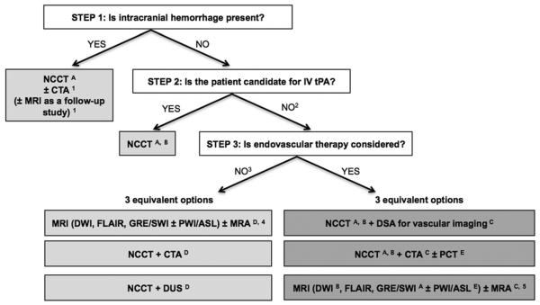
Suggested imaging protocols for patients presenting with acute stroke symptoms on the basis of the clinical scenario and the therapeutic options available. Each of the gray boxes represents one imaging strategy. So as not to delay treatment, a standardized imaging approach should be used: one imaging strategy (gray box) should be selected, and all imaging studies belonging to this strategy should be performed up front in as few sessions as possible. 1To assess the etiology of the intracranial hemorrhage (CT angiography [CTA] for vascular pathologies, such as aneurysms, arteriovenous malformations, vasculopathies; MRI for vascular malformations and neoplastic and other pathologies associated with hemorrhage). 2Also if the patient is not a candidate for intravenous (IV) tissue plasminogen activator (tPA) (contraindication to tPA, outside time window for tPA) or if IV tPA failed or it is thought that it might fail. 3For patients who are outside the time window for acute reperfusion therapies (>4.5 hours at sites at which only IV tPA is being considered; >8 hours at sites at which endovascular therapy is considered) and for patients with transient ischemic attacks (TIAs), emphasis is on secondary prevention, and their imaging workup should be focused on vascular imaging (CTA, MR angiography [MRA], or doppler ultrasound [DUS]) to assess the carotid arteries as a possible cause of the ischemic stroke, with secondary prevention in mind. If MRA is performed, it makes sense to concurrently obtain MRI with diffusion-weighted imaging (DWI), fluid-attenuated inversion recovery (FLAIR), and gradient-recalled echo (GRE) or susceptibility-weighted imaging (SWI). Echocardiography should also be performed to assess for cardiac sources. 4If available, MRI or MRA is the preferred imaging modality for patients with TIA. 5At institutions at which MRI is available 24/7 and can be performed within a short time after admission. A: to assess for intracranial hemorrhage; B: to assess the extent of ischemic core; C: to assess the location and extent of the intravascular clot; D: to assess carotid atherosclerotic disease; E: to assess the extent of viable tissue. ASL = arterial spin-labeled; NCCT = noncontrast CT; PCT = perfusion CT; PWI = perfusion-weighted imaging.
References
-
- Kidwell CS, Wintermark M. Imaging of intracranial haemorrhage. Lancet Neurol. 2008;7:256–67. - PubMed
-
- Huisman TA. Intracranial hemorrhage: ultrasound, CT and MRI findings. Eur Radiol. 2005;15:434–40. - PubMed
-
- Hoggard N, Wilkinson ID, Paley MN, Griffiths PD. Imaging of haemorrhagic stroke. Clin Radiol. 2002;57:957–68. - PubMed
-
- European Stroke Organisation (ESO) Executive Committee. ESO Writing Committee Guidelines for management of ischaemic stroke and transient ischaemic attack 2008. Cerebrovasc Dis. 2008;25:457–507. - PubMed
-
- Saver JL. Time is brain—quantified. Stroke. 2006;37:263–6. - PubMed
Publication types
MeSH terms
Grants and funding
LinkOut - more resources
Full Text Sources
Other Literature Sources
Medical
