. 2013;37(2):309-23.
doi: 10.3233/JAD-130182.

Blast exposure causes early and persistent aberrant phospho- and cleaved-tau expression in a murine model of mild blast-induced traumatic brain injury

Affiliations

Blast exposure causes early and persistent aberrant phospho- and cleaved-tau expression in a murine model of mild blast-induced traumatic brain injury

Bertrand R Huber et al. J Alzheimers Dis. 2013.

Abstract

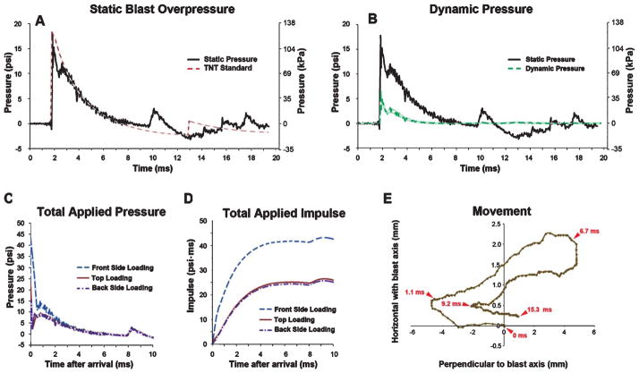
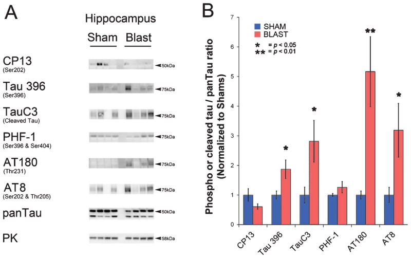
Mild traumatic brain injury (mTBI) is considered the 'signature injury' of combat veterans that have served during the wars in Iraq and Afghanistan. This prevalence of mTBI is due in part to the common exposure to high explosive blasts in combat zones. In addition to the threats of blunt impact trauma caused by flying objects and the head itself being propelled against objects, the primary blast overpressure (BOP) generated by high explosives is capable of injuring the brain. Compared to other means of causing TBI, the pathophysiology of mild-to-moderate BOP is less well understood. To study the consequences of BOP exposure in mice, we employed a well-established approach using a compressed gas-driven shock tube that recapitulates battlefield-relevant open-field BOP. We found that 24 hours post-blast a single mild BOP provoked elevation of multiple phospho- and cleaved-tau species in neurons, as well as elevating manganese superoxide-dismutase (MnSOD or SOD2) levels, a cellular response to oxidative stress. In hippocampus, aberrant tau species persisted for at least 30 days post-exposure, while SOD2 levels returned to sham control levels. These findings suggest that elevated phospho- and cleaved-tau species may be among the initiating pathologic processes induced by mild blast exposure. These findings may have important implications for efforts to prevent blast-induced insults to the brain from progressing into long-term neurodegenerative disease processes.


Figures

Fig. 1
Fig. 1
Shock tube: Photograph denotes the four main functional components of the shock tube: (i) the driver in which compressed helium is loaded. Helium was used rather than air because it is more easily adjusted to replicate the extremely short time course of the initial positive pressure pulse that is characteristic of high explosive detonations in the open-field; (ii) the spool assembly which is controlled by high-speed electronic valves that regulate simultaneous rupture of two mylar diaphragms at a pre-determined, highly reproducible psi (see Methods for description); (iii) the exposure area containing the animal restraint harness and anesthesia delivery system (see Methods for more details); and (iv) the attenuator which reduces reflections/rarefactions to improve correspondence of the shock waves with idealized open-field BOP characteristics (see Fig. 2) and which suppresses ambient noise to levels suitable for use in a biomedical research facility. Arrows denote placement of pressure gauges.
Fig. 2
Fig. 2
Blast overpressure characteristics and blast-induced head displacement. A) The time (ms) versus pressure (psi, left y-axis; kPa, right y-axis) plot shows an example of the static BOP used in this study as measured 5 cm directly above the animal (black trace) and illustrates the close correspondence of the shock tube-generated BOP with the Friedlander waveform expected from an open-field detonation of 15.9 kg TNT at 7.6m (dashed red trace). See Results for details. B) Green (dashed) trace indicates the intensity of the calculated dynamic pressure (blast wind) was approximately 3-fold less intense than the static BOP (solid black trace) is replotted from panel A. C) Illustrates total calculated applied pressure and (D) calculated total applied impulse (the impulse is the integration of pressure over the time of application) for BOP used in this study (A and B). E) Illustrates a representative mouse head excursion (determined from high speed video frame-by-frame analysis of a mark 2mm below eye).
Fig. 3
Fig. 3
Exploratory activity returned to normal yoked sham control levels within 24 hours after blast exposure. Histogram indicates mean beam crossings per min in activity cages normalized to shams measured 1 and 24 hours post exposure. One hour post-treatment BOP-exposed mice displayed significantly less exploratory behavior (p < 0.01) compared to sham controls. At 24 hours post-exposure activity among BOP-treated animals was comparable to sham controls (n = 5 BOP and n = 5 sham controls). Error bars indicate standard error of the mean (SEM).
Fig. 4
Fig. 4
Multiple pathologic phospho- and cleaved-tau species are increased 24 h after BOP. A) Representative western blots illustrate blast-induced elevation of phospho- and cleaved tau species in cortex, hippocampus, and cerebellum. Each lane corresponds to one sham or blast-exposed animal. B) Histograms indicate quantification of mean densitometric ratios for each tau epitope with respect to total pan-Tau immunoreactivity. The epitopes recognized by specific antibodies are denoted with reference to homologous phospho-amino acid sequences in human tau. Pyruvate kinase (PK) served as a gel protein load control. Indicated p-values are t-test results (n = 5 for blast and n = 5 for shams). Error bars indicate standard error of the means (SEM).
Fig. 5
Fig. 5
Blast exposure provokes increased pathologic tau and elevated SOD2 expression. A-B) Confocal microscopy revealed elevated CP13 immunoreactivity (red) in hippocampal CA4 pyramidal neurons of blast-exposed but not sham-treated mice. GFAP immunoreactivity in CA4 region (green) appeared comparable between BOP and sham animals. C, D) In cerebellum phospho-tau AT270 immunoreactivity was markedly increased in apparent basket cells of blast-exposed mice. Cell bodies stained with DAPI appear blue. E, F) Cleaved-tau immunostained by TauC3 (red) was evident in cerebellar Purkinje cell bodies of blast-exposed animals. G, H) SOD2 (green), a marker of mitochondrial oxidative stress, was markedly elevated in hippocampus of blast exposed animals. Overall hippocampal GFAP staining (purple) appeared comparable between sham and blast exposed animals. I, J) In cerebellum, Purkinje cells also displayed increasedSOD2expression. Blast-induced changes in GFAP levels (red) were less pronounced. K, L) GFAP immunoreactivity (green) in the glial limitans of parietal cortex and M, N) AT270 immunostaining (red) in cerebellar white matter tracks underlying the DAPI-stained granule cell layer was elevated in blast exposed animals. Scale bars indicate 40 m for panels A-F, I-L; 100 μm for G-H, M-N.
Fig. 6
Fig. 6
Increased SOD2 expression in hippocampus at 24 hours post-blast returned to sham levels with 30 days. A) At 24 hours post-exposure western blots revealed increased SOD2 levels in hippocampus of blast-exposed mice and by 30 days appeared comparable to control sham-treated animals. Each lane represents one BOP or sham animal. B) Histograms show quantification of mean densitometric SOD2 western blot immunoreactivty at 24 hours and 30 days post-treatment normalized with respect to yoked shams controls. Indicated p values are t-tests results (n = 5 BOP and n = 5 sham animals). Error bars indicate standard error of the means (SEM).
Fig. 7
Fig. 7
In hippocampus pathologic tau species were elevated 30 days following a single mild BOP. A) Representative western blots show that phospho-tau epitopes recognized by Tau396, AT180, and AT8 appear elevated in blast-exposed mice while total pan-tau immunoreactivity was comparable to sham animals. Cleaved tau recognized by TauC3 also appeared elevated. Pyruvate kinase (PK) served as a gel protein load control. B) Histograms indicated quantification of the ratios of phospho and cleaved tau with respect to total pan tau. Indicated p values are t-tests results (n = 5 BOP and n = 5 sham animals). Error bars indicate standard error of the means (SEM).

References

    1. Hosek B. How is deployment to Iraq and Afghanistan affecting U.S. service members and their families? An overview of early RAND research on the topic. RAND Corporation; Santa Monica California, USA: 2011. - PMC - PubMed
    1. Hoge CW, McGurk D, Thomas JL, Cox AL, Engel CC, Castro CA. Mild traumatic brain injury in U.S. Soldiers returning from Iraq. N Engl J Med. 2008;358:453–463. - PubMed
    1. McKee AC, Cantu RC, Nowinski CJ, Hedley-Whyte ET, Gavett BE, Budson AE, Santini VE, Lee HS, Kubilus CA, Stern RA. Chronic traumatic encephalopathy in athletes: Progressive tauopathy after repetitive head injury. J Neuropathol Exp Neurol. 2009;68:709–735. - PMC - PubMed
    1. Erlanger DM, Kutner KC, Barth JT, Barnes R. Neuropsychology of sports-related head injury: Dementia pugilistica to post concussion syndrome. Clin Neuropsychol. 1999;13:193–209. - PubMed
    1. Omalu BI, DeKosky ST, Minster RL, Kamboh MI, Hamilton RL, Wecht CH. Chronic traumatic encephalopathy in a National Football League player. Neurosurgery. 2005;57 :128–134. - PubMed

Publication types