

Global axonal transport rates are unaltered in htau mice in vivo
- PMID: 23948900
- PMCID: PMC3819434
- DOI: 10.3233/JAD-130671
Global axonal transport rates are unaltered in htau mice in vivo
Abstract
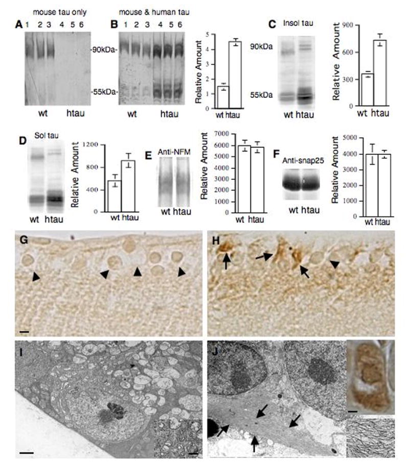
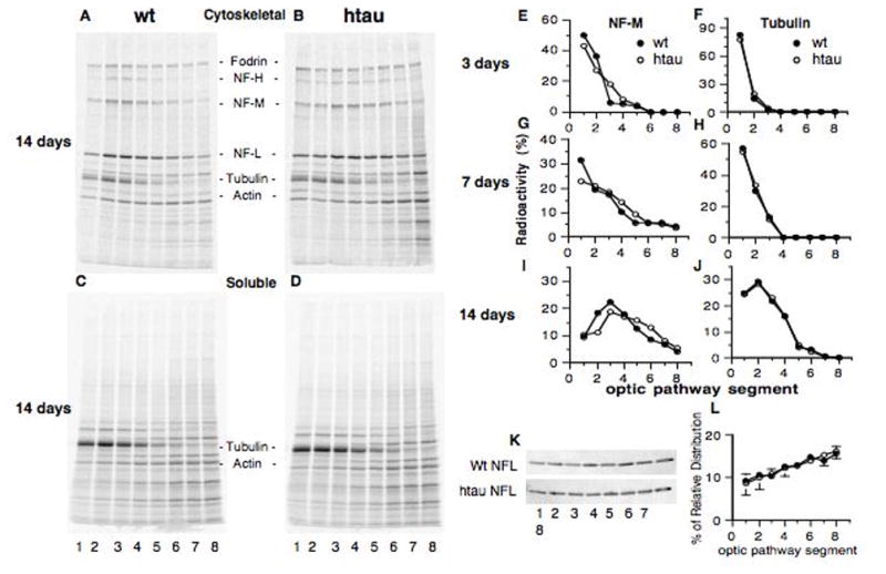
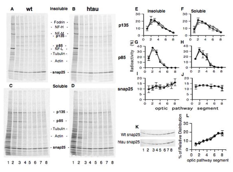
Microtubule-based axonal transport is believed to become globally disrupted in Alzheimer's disease in part due to alterations of tau expression or phosphorylation. We previously showed that axonal transport rates along retinal ganglion axons are unaffected by deletion of normal mouse tau or by overexpression of wild-type human tau. Here, we report that htau mice expressing 3-fold higher levels of human tau in the absence of mouse tau also display normal fast and slow transport kinetics despite the presence of abnormally hyperphosphorylated tau in some neurons. In addition, markers of slow transport (neurofilament light subunit) and fast transport (snap25) exhibit normal distributions along optic axons of these mice. These studies demonstrate that human tau overexpression, even when associated with a limited degree of tau pathology, does not necessarily impair general axonal transport function in vivo.
Figures
Similar articles
-
Axonal transport rates in vivo are unaffected by tau deletion or overexpression in mice.J Neurosci. 2008 Feb 13;28(7):1682-7. doi: 10.1523/JNEUROSCI.5242-07.2008. J Neurosci. 2008. PMID: 18272688 Free PMC article.
-
Tau accumulation in the retina promotes early neuronal dysfunction and precedes brain pathology in a mouse model of Alzheimer's disease.Mol Neurodegener. 2017 Aug 3;12(1):58. doi: 10.1186/s13024-017-0199-3. Mol Neurodegener. 2017. PMID: 28774322 Free PMC article.
-
In vivo axonal transport deficits in a mouse model of fronto-temporal dementia.Neuroimage Clin. 2014 Mar 31;4:711-7. doi: 10.1016/j.nicl.2014.02.005. eCollection 2014. Neuroimage Clin. 2014. PMID: 24936422 Free PMC article.
-
Axonal transport, tau protein, and neurodegeneration in Alzheimer's disease.Neuromolecular Med. 2002;2(2):151-65. doi: 10.1385/NMM:2:2:151. Neuromolecular Med. 2002. PMID: 12428809 Review.
-
Tau and Axonal Transport Misregulation in Tauopathies.Adv Exp Med Biol. 2019;1184:81-95. doi: 10.1007/978-981-32-9358-8_7. Adv Exp Med Biol. 2019. PMID: 32096030 Free PMC article. Review.
Cited by
-
Motor and cognitive deficits in aged tau knockout mice in two background strains.Mol Neurodegener. 2014 Aug 14;9:29. doi: 10.1186/1750-1326-9-29. Mol Neurodegener. 2014. PMID: 25124182 Free PMC article.
-
Tau Isoforms Imbalance Impairs the Axonal Transport of the Amyloid Precursor Protein in Human Neurons.J Neurosci. 2017 Jan 4;37(1):58-69. doi: 10.1523/JNEUROSCI.2305-16.2016. J Neurosci. 2017. PMID: 28053030 Free PMC article.
-
Differential Hyperphosphorylation of Tau-S199, -T231 and -S396 in Organotypic Brain Slices of Alzheimer Mice. A Model to Study Early Tau Hyperphosphorylation Using Okadaic Acid.Front Aging Neurosci. 2018 Apr 19;10:113. doi: 10.3389/fnagi.2018.00113. eCollection 2018. Front Aging Neurosci. 2018. PMID: 29725295 Free PMC article.
-
Axonal transport defects in Alzheimer's disease.Mol Neurobiol. 2015;51(3):1309-21. doi: 10.1007/s12035-014-8810-x. Epub 2014 Jul 23. Mol Neurobiol. 2015. PMID: 25052480 Review.
-
Intracellular amyloid β oligomers impair organelle transport and induce dendritic spine loss in primary neurons.Acta Neuropathol Commun. 2015 Aug 21;3:51. doi: 10.1186/s40478-015-0230-2. Acta Neuropathol Commun. 2015. PMID: 26293809 Free PMC article.
References
-
- Ballatore C, Lee VM, Trojanowski JQ. Tau-mediated neurodegeneration in Alzheimer’s disease and related disorders. Nat Rev Neurosci. 2007;8:663–672. - PubMed
-
- Caceres A, Kosik KS. Inhibition of neurite polarity by tau antisense oligonucleotides in primary cerebellar neurons. Nature. 1990;343:461–463. - PubMed
-
- Gomez-Isla T, Hollister R, West H, Mui S, Growdon JH, Petersen RC, Parisi JE, Hyman BT. Neuronal loss correlates with but exceeds neurofibrillary tangles in Alzheimer’s disease. Ann Neurol. 1997;41:17–24. - PubMed
Publication types
MeSH terms
Substances
Grants and funding
LinkOut - more resources
Full Text Sources
Other Literature Sources
Molecular Biology Databases
