

Varus thrust and knee frontal plane dynamic motion in persons with knee osteoarthritis
- PMID: 23948980
- PMCID: PMC4014355
- DOI: 10.1016/j.joca.2013.08.007
Varus thrust and knee frontal plane dynamic motion in persons with knee osteoarthritis
Abstract
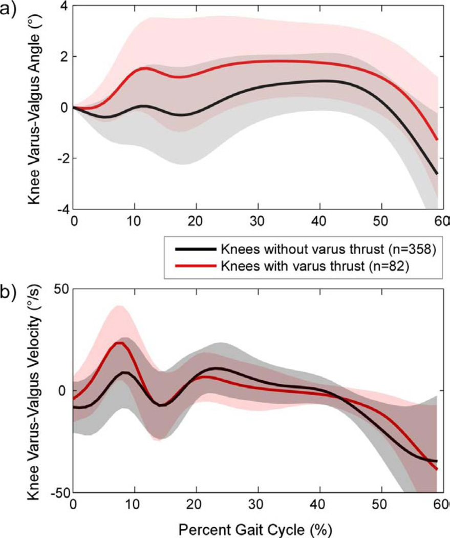
Objective: Varus thrust visualized during walking is associated with a greater medial knee load and an increased risk of medial knee osteoarthritis (OA) progression. Little is known about how varus thrust presence determined by visual observation relates to quantitative gait kinematic data. We hypothesized that varus thrust presence is associated with greater knee frontal plane dynamic movement during the stance phase of gait.
Methods: Participants had knee OA in at least one knee. Trained examiners assessed participants for varus thrust presence during ambulation. Frontal plane knee motion during ambulation was captured using external passive reflective markers and an 8-camera motion analysis system. To examine the cross-sectional relationship between varus thrust and frontal plane knee motion, we used multivariable regression models with the quantitative motion measures as dependent variables and varus thrust (present/absent) as predictor; models were adjusted for age, gender, body mass index (BMI), gait speed, and knee static alignment.
Results: 236 persons [mean BMI: 28.5 kg/m(2) (standard deviation (SD) 5.5), mean age: 64.9 years (SD 10.4), 75.8% women] contributing 440 knees comprised the study sample. 82 knees (18.6%) had definite varus thrust. Knees with varus thrust had greater peak varus angle and greater peak varus angular velocity during stance than knees without varus thrust (mean differences 0.90° and 6.65°/s, respectively). These patterns remained significant after adjusting for age, gender, BMI, gait speed, and knee static alignment.
Conclusion: Visualized varus thrust during walking was associated with a greater peak knee varus angular velocity and a greater peak knee varus angle during stance phase of gait.
Keywords: Gait analysis; Instability; Knee osteoarthritis; Varus thrust.
Copyright © 2013 Osteoarthritis Research Society International. Published by Elsevier Ltd. All rights reserved.
Figures
References
-
- Schipplein OD, Andriacchi TP. Interaction between active and passive knee stabilizers during level walking. J Orthop Res. 1991;9(1):113–119. - PubMed
-
- Andriacchi TP, Mündermann A, Smith RL, Alexander EJ, Dyrby CO, Koo S. A framework for the in vivo pathomechanics of osteoarthritis at the knee. Ann Biomed Eng. 2004;32(3):447–457. - PubMed
-
- Bennell KL, Bowles K-A, Wang Y, Cicuttini F, Davies-Tuck M, Hinman RS. Higher dynamic medial knee load predicts greater cartilage loss over 12 months in medial knee osteoarthritis. Ann Rheum Dis. 2011;70(10):1770–1774. - PubMed
-
- Chang A, Hayes K, Dunlop D, Hurwitz D, Song J, Cahue S, et al. Thrust during ambulation and the progression of knee osteoarthritis. Arthritis Rheum. 2004;50(12):3897–3903. - PubMed
Publication types
MeSH terms
Grants and funding
LinkOut - more resources
Full Text Sources
Other Literature Sources
