. 2013 Aug 15;4(8):e773.
doi: 10.1038/cddis.2013.279.

KCa3.1 channels are involved in the infiltrative behavior of glioblastoma in vivo

Affiliations

KCa3.1 channels are involved in the infiltrative behavior of glioblastoma in vivo

G D'Alessandro et al. Cell Death Dis. .

Abstract

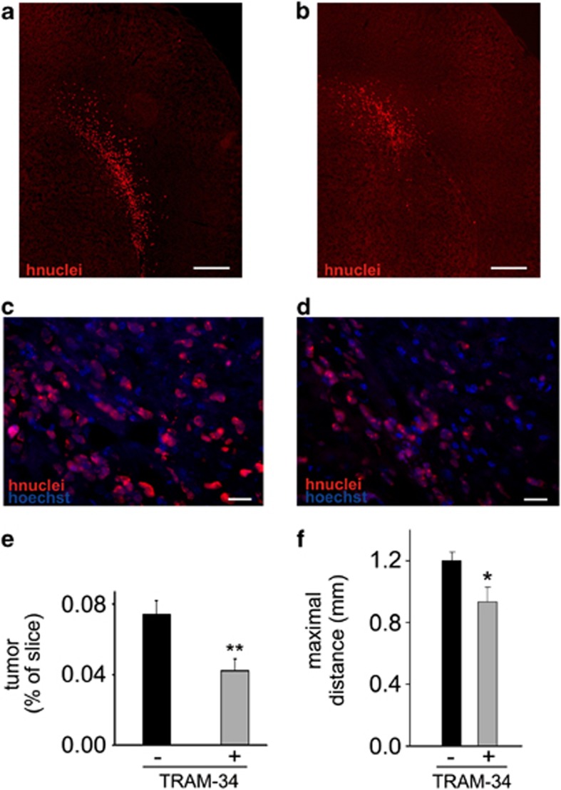
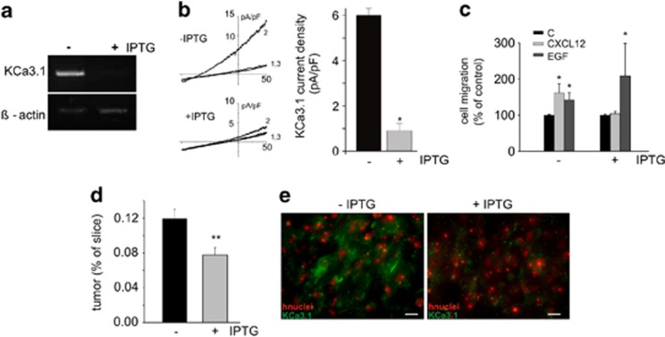
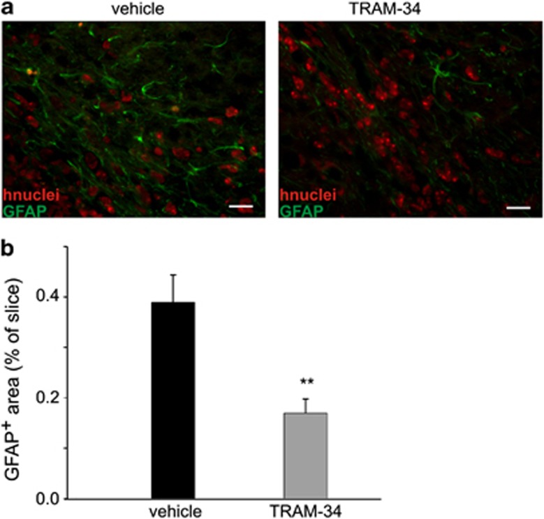
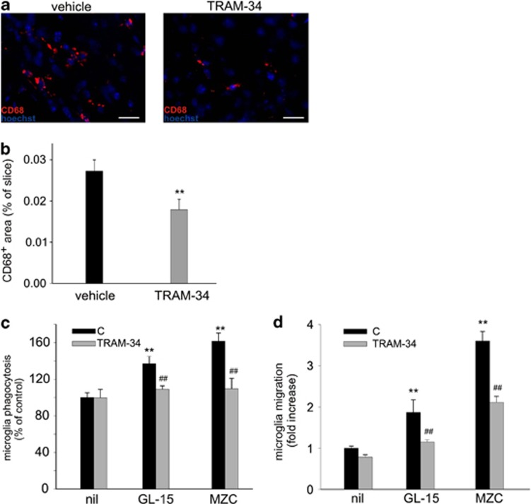
Glioblastoma multiforme (GBM) is a diffuse brain tumor characterized by high infiltration in the brain parenchyma rendering the tumor difficult to eradicate by neurosurgery. Efforts to identify molecular targets involved in the invasive behavior of GBM suggested ion channel inhibition as a promising therapeutic approach. To determine if the Ca(2+)-dependent K(+) channel KCa3.1 could represent a key element for GBM brain infiltration, human GL-15 cells were xenografted into the brain of SCID mice that were then treated with the specific KCa3.1 blocker TRAM-34 (1-((2-chlorophenyl) (diphenyl)methyl)-1H-pyrazole). After 5 weeks of treatment, immunofluorescence analyses of cerebral slices revealed reduced tumor infiltration and astrogliosis surrounding the tumor, compared with untreated mice. Significant reduction of tumor infiltration was also observed in the brain of mice transplanted with KCa3.1-silenced GL-15 cells, indicating a direct effect of TRAM-34 on GBM-expressed KCa3.1 channels. As KCa3.1 channels are also expressed on microglia, we investigated the effects of TRAM-34 on microglia activation in GL-15 transplanted mice and found a reduction of CD68 staining in treated mice. Similar results were observed in vitro where TRAM-34 reduced both phagocytosis and chemotactic activity of primary microglia exposed to GBM-conditioned medium. Taken together, these results indicate that KCa3.1 activity has an important role in GBM invasiveness in vivo and that its inhibition directly affects glioma cell migration and reduces astrocytosis and microglia activation in response to tumor-released factors. KCa3.1 channel inhibition therefore constitutes a potential novel therapeutic approach to reduce GBM spreading into the surrounding tissue.


Figures

Figure 1
Figure 1
TRAM-34 reduces tumor-infiltrated area and maximal migration distance of GL-15 in the mouse brain. Brain slices stained with anti human nuclei (hnuclei, in red) in vehicle- (a, c) and TRAM-34- (b, d) treated mice. a and b show the infiltrated (right) hemisphere ( × 10); c and d are magnifications ( × 40; total nuclei: blue, Hoechst) of a part of the areas indicated in a and b; (e) quantification of tumor-infiltrated area (area occupied by human nuclei-positive cells versus total slice area) in vehicle- and TRAM-34-treated mice (**P<0.001, Student's t-test). (f) brain slices were analyzed for the presence of GL-15 cells (cells positive for human nuclei staining) in the sagittal plane. Values are the maximal distances (in mm) obtained in vehicle- (n=6) and TRAM-34-treated mice (n=6). *P<0.05, Student's t-test. Bars: a, b: 225 μm; c, d: 10 μm
Figure 2
Figure 2
KCa3.1 shRNA-silenced GL-15 cells show reduced infiltration in the mouse brain. (a) mRNA quantification of KCa3.1 relative to β-actin in control and IPTG-induced cells; (b) left, representative I–Vs from control and IPTG-induced cells, in C (1), NS309 (10 μM) (2), and NS309/TRAM-34 (3 μM) (3). Right, bar plot showing the TRAM-34-sensitive KCa3.1 current density in control and IPTG-induced cells *P<0.05; (c) control and IPTG-treated cell migration toward CXCL12 or EGF (*P<0.05 versus C of the same experimental group, Student's t-test); (d) quantification of tumor-infiltrated area in the brain of mice injected with shRNA-silenced GL-15 cells (area occupied by human nuclei-positive cells versus total slice area) and treated or not with IPTG (**P<0.01, Student's t-test); (e) immunofluorescence analysis of KCa3.1 expression (green; hnuclei: red) in brain slices obtained from mice injected with shRNA-silenced GL-15 cells un-induced (left) or induced (right) with IPTG. Bars, 10 μm
Figure 3
Figure 3
TRAM-34 reduces astrogliosis. (a) representative immunofluorescences of GFAP (green) and human nuclei (red) in vehicle- and TRAM-34- treated mice ( × 40; bars 10 μm); (b) quantification of astrogliosis (area occupied by GFAP-positive cells versus total slice area) in vehicle- and TRAM-34-treated mice (**P<0.001, Student's t-test)
Figure 4
Figure 4
TRAM-34 treatment reduces microglia activation and migration toward GBM. Brain slices obtained from mice xenografted with GL-15 GBM were analyzed for CD68 immunoreactivity (red; total nuclei: blue, Hoechst) in vehicle- and TRAM-34-treated mice (a, bars 10 μm). Mean values of CD68 staining per slice are reported in (b). Data are expressed as the ratio between the area occupied by CD68 positive cells versus total slice area and are the mean of 12 slices per mouse (n=4) for each condition. (c) In vitro phagocytosis of fluorescent FluoSpheres by microglia exposed for 18 h to control medium (nil) or media conditioned (24 h) by GL-15 and MZC cells in the presence or in the absence of TRAM-34 (5 μM). Only cells phagocyting at least three beads were scored as positive and results are expressed as % of control (n=3–5; **P<0.001). (d) In vitro migration of microglia toward control medium (nil) or media conditioned (24 h) by GL-15 and MZC cells in the presence or in the absence of TRAM-34 (5 μM). Migration is expressed as fold increase in the number of migrating cells in comparison with C, nil-exposed cultures and results are the mean±S.E.M. of 5–6 independent experiments

References

    1. Giese A, Bjerkvig R, Berens ME, Westphal M. Cost of migration: invasion of malignant gliomas and implications for treatment. J Clin Oncol. 2003;21:1624–1636. - PubMed
    1. Wen PY, Kesari S. Malignant gliomas in adults. N Engl J Med. 2008;359:492–507. - PubMed
    1. Markovic DS, Glass R, Synowitz M, Nv Rooijen, Kettenmann H. Microglia stimulate the invasiveness of glioma cells by increasing the activity of metalloprotease-2. J Neuropathol Exp Neurol. 2005;64:754–762. - PubMed
    1. Wesolowska A, Kwiatkowska A, Slomnicki L, Dembinski M, Master A, Sliwa M, et al. Microglia-derived TGF-beta as an important regulator of glioblastoma invasion-an inhibition of TGF-beta-dependent effects by shRNA against human TGF-beta type II receptor. Oncogene. 2008;27:918–930. - PubMed
    1. Charles NA, Holland EC, Gilbertson R, Glass R, Kettenmann H. The brain tumor microenvironment. Glia. 2012;60:502–514. - PubMed

Publication types

MeSH terms