Review
. 2013 Sep;14(9):645-60.
doi: 10.1038/nrg3521.

New genes as drivers of phenotypic evolution

Affiliations
Review

New genes as drivers of phenotypic evolution

Sidi Chen et al. Nat Rev Genet. 2013 Sep.

Erratum in

  • Nat Rev Genet. 2013 Oct;14(10):744

Abstract

During the course of evolution, genomes acquire novel genetic elements as sources of functional and phenotypic diversity, including new genes that originated in recent evolution. In the past few years, substantial progress has been made in understanding the evolution and phenotypic effects of new genes. In particular, an emerging picture is that new genes, despite being present in the genomes of only a subset of species, can rapidly evolve indispensable roles in fundamental biological processes, including development, reproduction, brain function and behaviour. The molecular underpinnings of how new genes can develop these roles are starting to be characterized. These recent discoveries yield fresh insights into our broad understanding of biological diversity at refined resolution.


Conflict of interest statement

Competing interests statement

The authors declare no competing financial interests.

Figures
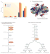
Figure 1
Figure 1. Several new genes evolved novel biochemical functions
a | The protein Jingwei (Jgw) encoded by African Drosophila spp. is a dehydrogenase enzyme that has evolved altered substrate specificity compared with the ancestral Alcohol dehydrogenase (Adh). Compared with the parental enzyme, Jgw can use long-chain primary alcohols more efficiently; in particular, Jgw has greater activities than the ancestral Adh towards farnesol (which is involved in juvenile hormone biosynthesis) and geraniol (which is involved in recruitment pheromone biosynthesis). b | The locations of the substitutions on the structure of the dimeric Adh. c,d | Three recent duplicates in the cytochrome P450 family in Arabidopsis thaliana led to the assembly of a new pathway of N1,N5-di(hydroxyferuloyl)-N10-sinapoyl spermidine biosynthesis (the new duplicates CYP98A8 and CYP98A9 are shown in red) (part c) and a pathway for arabidopyrone biosynthesis (the young duplicated enzyme CYP84A4 is shown in red) (part d). D. teissieri, Drosophila teissieri; D. yakuba, Drosophila yakuba. Parts a and b are modified, with permission, from REF. © (2004) US National Academy of Sciences.
Figure 2
Figure 2. New genes participate in developmental processes in Drosophila spp
a,b | The experimental procedures (part a) assessed the developmental importance of the new genes that have originated during Drosophila spp. speciation (as mapped in the evolutionary tree by comparing the genomes of different Drosophila spp. (part b)). The values in the tree indicate the numbers of new genes in the Drosophila melanogaster (D. mel) lineage at each evolutionary branchpoint; 566 genes were chosen for designing knockdown experiments from the total of 727 new genes that have originated since branch 1 in the tree. c,d | A summary of the main experimental results from the 195 new genes tested for the effect on development following gene knockdown. First, knockdown of approximately one-third of either new genes or old genes resulted in lethality, indicating that the proportion of essential new genes is similar to the proportion of essential old genes in branch 0 of the evolutionary tree (part c). Second, knockdown of older genes led to the termination of development at earlier stages than knockdown of new genes, indicating that old genes function earlier in development than new genes, most of which have essential functions in the pupal stages (part d). D. ana, Drosophila ananassae; D. ere, Drosophila erecta; D. gri, Drosophila grimshawi; D. mel, Drosophila melanogaster; D. moj, Drosophila mojavensis; D. per, Drosophila persimilis; D. pse, pseudoobscura; D. sec, Drosophila sechellia; D. sim, Drosophila simulans; D. wil, Drosophila willistoni; D. vir, Drosophila virilise; D. yak, Drosophila yakuba.
Figure 3
Figure 3. The involvement of new genes in the development of Drosophila spp., and in the brains of Drosophila spp. and humans
a | The phylogeny shows the distributions of new Drosophila spp. genes involved in development (above) and in the brain (below) in various evolutionary stages within the past 36 million years. The numbers in the ovals show the divergence time (in millions of years) between Drosophila melanogaster and various Drosophila spp. Red represents the number of new genes that were found to have essential developmental functions, whereas blue shows the number of new genes that were non-essential in development. Green represents the number of new genes that were expressed in the brain, and yellow shows the number of the non-brain-expressed new genes in reverse transcription PCR screening experiments. The left-most pie charts show the total numbers of new genes across all analysed stages. For the developmental data, the origination events at 3–6 million years ago (MYA) and 0–3 MYA are pooled. These data reveal that older genes are no more likely than newer genes to have evolved essential developmental functions or brain expression, suggesting the rapid evolution of phenotypic effects in new genes. b | The phylogeny shows the numbers of new genes in the human genome that originated at various stages during the divergence of human ancestors from the ancestors of other primates through duplication (DNA based and RNA based) and de novo mechanisms (BOX 1). The numbers within the ovals show divergence time (MYA). The numbers in the denominators are the total numbers of new genes that originated in each evolutionary branch, as identified previously, and the red numerators are the numbers of those new genes that are expressed in the prefrontal cortex (PFC), as detected based on the available microarray expression data. The PFC is the anterior part of the frontal lobe of the neocortex, which is implicated in cognitive functions in the developing human brain. These data suggest that new genes have been frequently acquired into the PFC transcriptome. c | The SLIT–ROBO RHO GTPase-activating protein 2C (SRGAP2C) gene is one of the 54 PFC-expressed human-specific new genes (the end branch of the tree in part b). It was formed by DNA-based duplication and has been subjected to extensive genetic and evolutionary analyses, and functional characterization. The gene structure shows that SRGAP2C (bottom) is a duplicate of the amino-terminal part of the parental gene, SRGAP2 (top). The transgenic expression of SRGAP2C in cultured mouse cortical neurons induces denser and longer spines, as shown in the dendrite images (scale bar represents 2 μm), and the measured spine density and neck length, shown in the graphs on the right. *** indicates a significance of P<0.001. D. ananassae, Drosophila ananassae; D. pseudoobscura, Drosophila pseudoobscura; D. simulans, Drosophila simulans; D. willistoni, Drosophila willistoni; D. yakuba, Drosophila yakuba. Part c is modified, with permission, from REF. © (2012) Elsevier Science.
Figure 4
Figure 4. The functions of the young nsr gene in sperm development in Drosophila spp
a | The novel spermatogenesis regulator (nsr) gene was formed through the DNA-based duplication 3–6 million years ago (MYA) in the common ancestor of the clade including Drosophila melanogaster, Drosophila simulans and Drosophila sechellia. The two outgroups shown are Drosophila ananassae (which separated 11 MYA) and Drosophila yakuba (which separated 6 MYA). b | A schematic cross-section of a normal sperm axoneme with wild-type nsr: the outer (pink) and inner (blue) dynein arms are bounded by nine doublet microtubules (green circles) that surround a central pair of singlet microtubules (yellow circles) through radial spokes (grey). c,d | The functional importance of nsr was shown by P-element-mediated homozygous inactivation of nsr (nsr−/−). As seen from the electron micrographs, inactivation of nsr (part d) results in the loss of the outer dynein arms of the axoneme (white arrows) compared with the wild type (part c), whereas the inner arms remain normal (black arrows); scale bars represent 50 nm. The consequences in nsr−/− sperm are coiled axonemes and deficiencies in individualization. Figure is modified from REF. .
Figure 5
Figure 5. The rapid and extensive evolution of gene expression networks by integration of the new gene Zeus in Drosophila spp
a | Zeus, a new gene derived from the retrotransposition of the parental Caf40 gene, evolved under positive selection, as detected by the McDonald–Kreitman test and by the Ka/Ks ratio (defined as the ratio of the number of substitutions at non-synonymous sites to the number of substitutions at synonymous sites). In contrast to the slow evolution of Caf40 (blue), Zeus evolved rapidly in its protein sequences (red). The ratios displayed throughout the tree are Ka/Ks ratios, except those in red, which show the ratio of the number of non-synonymous sites to the number of synonymous polymorphic sites of nucleotides in the alleles of the natural population in Drosophila melanogaster and Drosophila simulans. b | The left panels show a comparison of the DNA-binding sites of Caf40 (blue) and Zeus (Red), as determined by chromatin immunoprecipitation followed by microarray (ChIP–chip). Separate analyses revealed that almost all of these sites have protein-coding genes downstream (not shown). The right panels show the genes for which expression correlates with the expression of Zeus, as determined by high-throughput RNA sequencing (RNA-seq) following RNA interference (RNAi)-mediated knockdown of Zeus, and the raw data is shown in the heatmap. The extensive changes in DNA-binding sites revealed that the integration of new genes reshaped the gene expression networks. D. ananassae, Drosophila ananassae; D. yakuba, Drosophila yakuba; IP, immunoprecipitation. Figure is modified, with permission, from REF. © (2012) Macmillan Publishers Ltd. All rights reserved.

References

    1. Long M, Betran E, Thornton K, Wang W. The origin of new genes: glimpses from the young and old. Nature Rev Genet. 2003;4:865–875. - PubMed
    1. Kaessmann H. Origins, evolution, and phenotypic impact of new genes. Genome Res. 2010;20:1313–1326. - PMC - PubMed
    1. Ding Y, Zhou Q, Wang W. Origins of new genes and evolution of their novel functions. Annu Rev Ecol Evol Syst. 2012;43:345–363.
    1. Long M, VanKuren NW, Chen S, Vibranovski MD. New gene evolution: little did we know. Annu Rev Genet. (in the press) - PMC - PubMed
    1. Hardison RC. Evolution of hemoglobin and its genes. Cold Spring Harb Perspect Med. 2012;2:a011627. - PMC - PubMed

Publication types