MicroRNA-155 drives TH17 immune response and tissue injury in experimental crescentic GN
- PMID: 23949802
- PMCID: PMC3839549
- DOI: 10.1681/ASN.2013020130
MicroRNA-155 drives TH17 immune response and tissue injury in experimental crescentic GN
Abstract
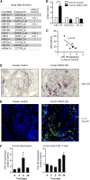
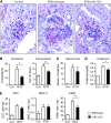
CD4(+) T cells play a pivotal role in the pathogenesis of autoimmune disease, including human and experimental crescentic GN. Micro-RNAs (miRs) have emerged as important regulators of immune cell development, but the impact of miRs on the regulation of the CD4(+) T cell immune response remains to be fully clarified. Here, we report that miR-155 expression is upregulated in the kidneys of patients with ANCA-associated crescentic GN and a murine model of crescentic GN (nephrotoxic nephritis). To elucidate the potential role of miR-155 in T cell-mediated inflammation, nephritis was induced in miR-155(-/-) and wild-type mice. The systemic and renal nephritogenic TH17 immune response decreased markedly in nephritic miR-155(-/-) mice. Consistent with this finding, miR-155-deficient mice developed less severe nephritis, with reduced histologic and functional injury. Adoptive transfer of miR-155(-/-) and wild-type CD4(+) T cells into nephritic recombination activating gene 1-deficient (Rag-1(-/-)) mice showed the T cell-intrinsic importance of miR-155 for the stability of pathogenic TH17 immunity. These findings indicate that miR-155 drives the TH17 immune response and tissue injury in experimental crescentic GN and show that miR-155 is a potential therapeutic target in TH17-mediated diseases.
Figures


Comment in
-
MicroRNA-155 a new therapeutic target in crescentic GN.J Am Soc Nephrol. 2013 Dec;24(12):1927-9. doi: 10.1681/ASN.2013090924. Epub 2013 Oct 24. J Am Soc Nephrol. 2013. PMID: 24158984 Free PMC article. No abstract available.
Similar articles
-
IL-17C/IL-17 Receptor E Signaling in CD4+ T Cells Promotes TH17 Cell-Driven Glomerular Inflammation.J Am Soc Nephrol. 2018 Apr;29(4):1210-1222. doi: 10.1681/ASN.2017090949. Epub 2018 Feb 26. J Am Soc Nephrol. 2018. PMID: 29483158 Free PMC article.
-
IL-17F Promotes Tissue Injury in Autoimmune Kidney Diseases.J Am Soc Nephrol. 2016 Dec;27(12):3666-3677. doi: 10.1681/ASN.2015101077. Epub 2016 Mar 30. J Am Soc Nephrol. 2016. PMID: 27030744 Free PMC article.
-
IL-17 Receptor C Signaling Controls CD4+ TH17 Immune Responses and Tissue Injury in Immune-Mediated Kidney Diseases.J Am Soc Nephrol. 2021 Dec;32(12):3081-3098. doi: 10.1681/ASN.2021030426. J Am Soc Nephrol. 2021. PMID: 35167487 Free PMC article.
-
Plasticity and heterogeneity of Th17 in immune-mediated kidney diseases.J Autoimmun. 2018 Feb;87:61-68. doi: 10.1016/j.jaut.2017.12.005. Epub 2017 Dec 21. J Autoimmun. 2018. PMID: 29275837 Review.
-
Mechanisms and functions of IL-17 signaling in renal autoimmune diseases.Mol Immunol. 2018 Dec;104:90-99. doi: 10.1016/j.molimm.2018.09.005. Epub 2018 Nov 15. Mol Immunol. 2018. PMID: 30448610 Review.
Cited by
-
T cell plasticity in renal autoimmune disease.Cell Tissue Res. 2021 Aug;385(2):323-333. doi: 10.1007/s00441-021-03466-z. Epub 2021 May 3. Cell Tissue Res. 2021. PMID: 33937944 Free PMC article. Review.
-
miR-34c attenuates epithelial-mesenchymal transition and kidney fibrosis with ureteral obstruction.Sci Rep. 2014 Apr 3;4:4578. doi: 10.1038/srep04578. Sci Rep. 2014. PMID: 24694752 Free PMC article.
-
Could IL-17A Be a Novel Therapeutic Target in Diabetic Nephropathy?J Clin Med. 2020 Jan 19;9(1):272. doi: 10.3390/jcm9010272. J Clin Med. 2020. PMID: 31963845 Free PMC article. Review.
-
Circulating miRNAs correlate with clinical evaluation of activity in ANCA-associated glomerulonephritis.Eur J Med Res. 2025 Jul 21;30(1):648. doi: 10.1186/s40001-025-02905-9. Eur J Med Res. 2025. PMID: 40685338 Free PMC article.
-
Renal Tissue miRNA Expression Profiles in ANCA-Associated Vasculitis-A Comparative Analysis.Int J Mol Sci. 2021 Dec 22;23(1):105. doi: 10.3390/ijms23010105. Int J Mol Sci. 2021. PMID: 35008531 Free PMC article.
References
-
- Tipping PG, Holdsworth SR: T cells in crescentic glomerulonephritis. J Am Soc Nephrol 17: 1253–1263, 2006 - PubMed
-
- Chadban SJ, Atkins RC: Glomerulonephritis. Lancet 365: 1797–1806, 2005 - PubMed
-
- Couser WG: Basic and translational concepts of immune-mediated glomerular diseases. J Am Soc Nephrol 23: 381–399, 2012 - PubMed
-
- Mosmann TR, Cherwinski H, Bond MW, Giedlin MA, Coffman RL: Two types of murine helper T cell clone. I. Definition according to profiles of lymphokine activities and secreted proteins. J Immunol 136: 2348–2357, 1986 - PubMed
-
- Harrington LE, Hatton RD, Mangan PR, Turner H, Murphy TL, Murphy KM, Weaver CT: Interleukin 17-producing CD4+ effector T cells develop via a lineage distinct from the T helper type 1 and 2 lineages. Nat Immunol 6: 1123–1132, 2005 - PubMed
Publication types
MeSH terms
Substances
LinkOut - more resources
Full Text Sources
Other Literature Sources
Molecular Biology Databases
Research Materials

