. 2013;9(8):e1003539.
doi: 10.1371/journal.ppat.1003539. Epub 2013 Aug 8.

SIGIRR, a negative regulator of TLR/IL-1R signalling promotes Microbiota dependent resistance to colonization by enteric bacterial pathogens

Affiliations

SIGIRR, a negative regulator of TLR/IL-1R signalling promotes Microbiota dependent resistance to colonization by enteric bacterial pathogens

Ho Pan Sham et al. PLoS Pathog. 2013.

Abstract

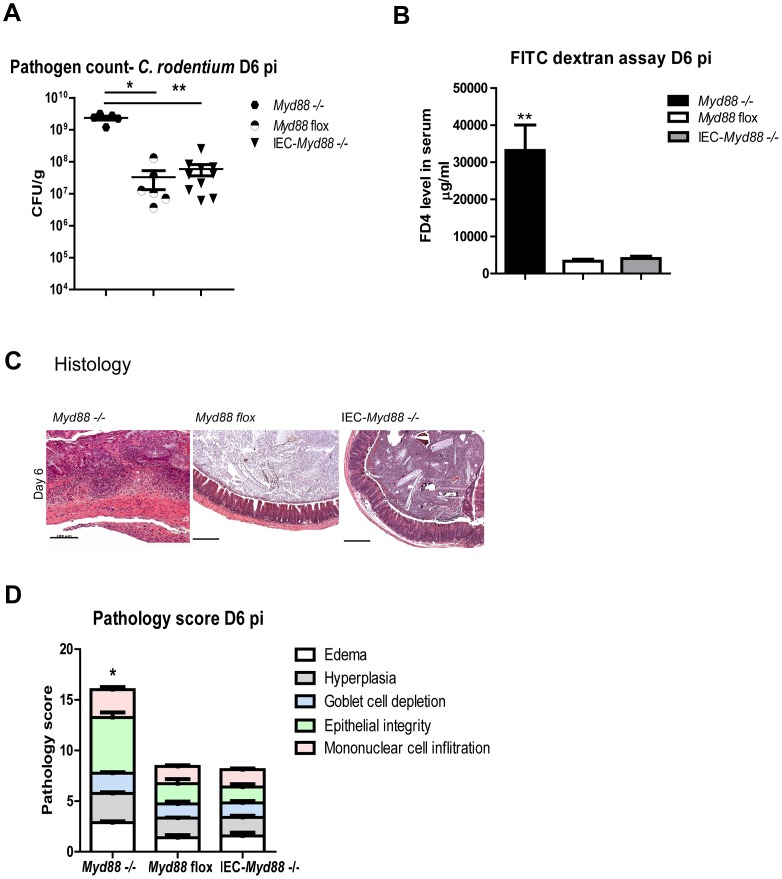
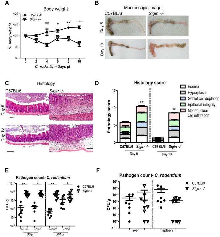
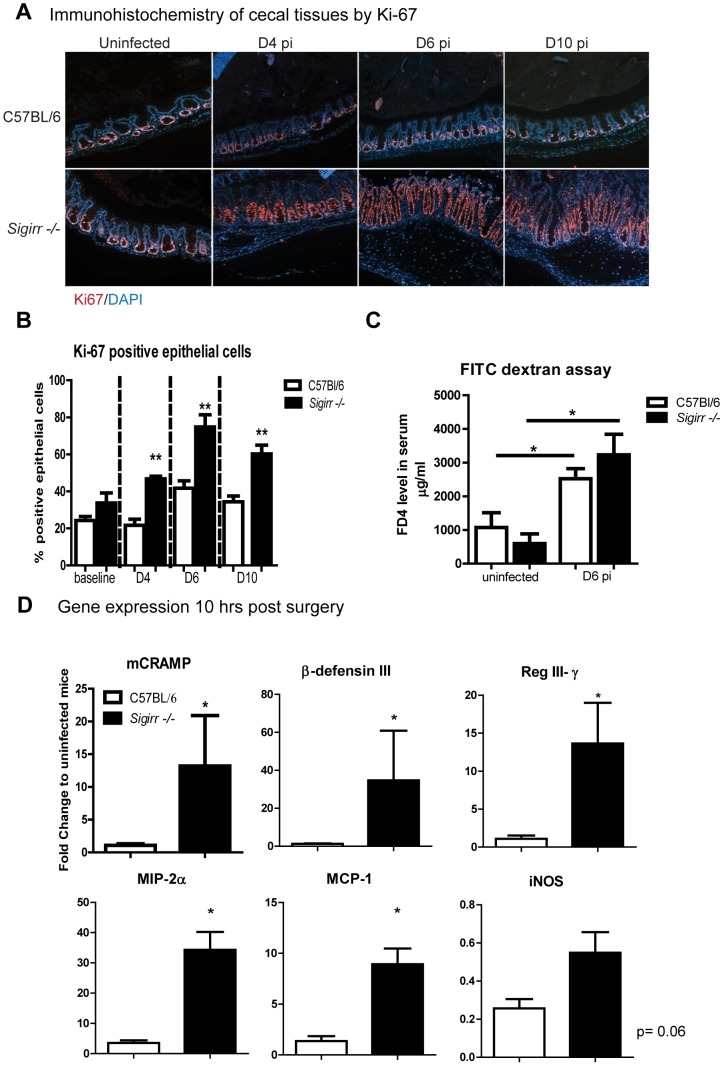
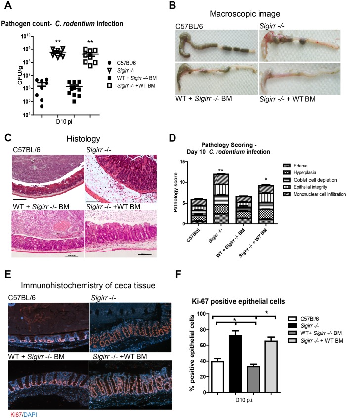
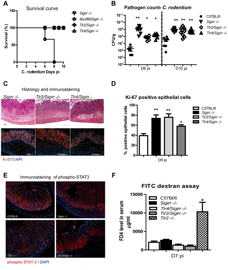
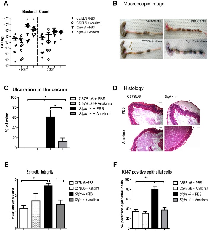
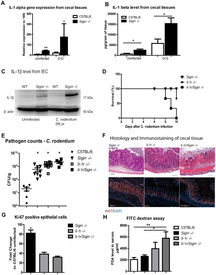
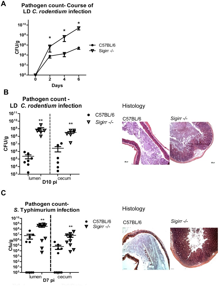
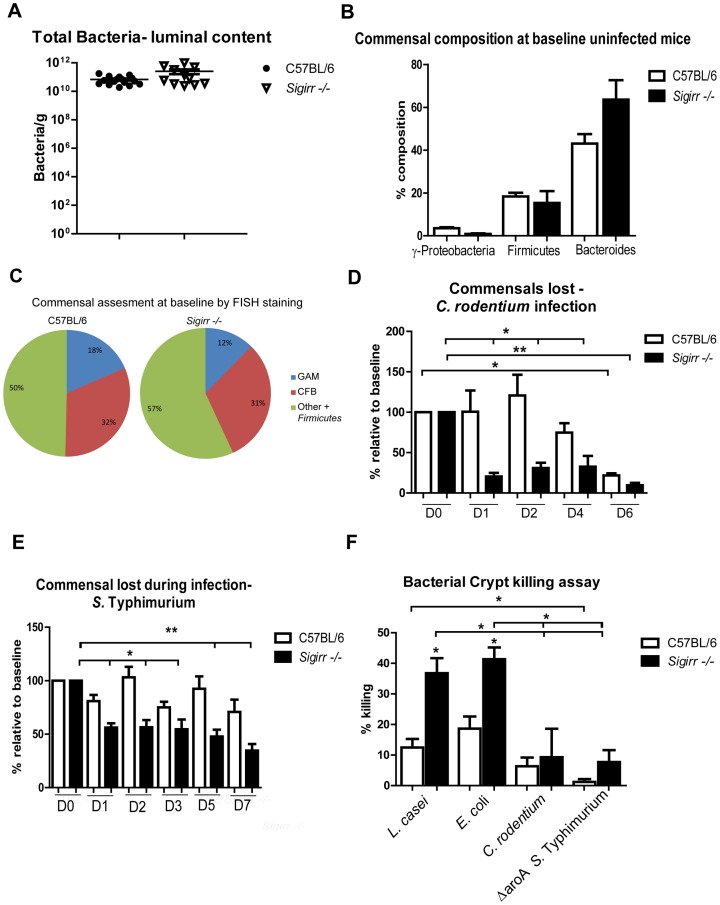
Enteric bacterial pathogens such as enterohemorrhagic E. coli (EHEC) and Salmonella Typhimurium target the intestinal epithelial cells (IEC) lining the mammalian gastrointestinal tract. Despite expressing innate Toll-like receptors (TLRs), IEC are innately hypo-responsive to most bacterial products. This is thought to prevent maladaptive inflammatory responses against commensal bacteria, but it also limits antimicrobial responses by IEC to invading bacterial pathogens, potentially increasing host susceptibility to infection. One reason for the innate hypo-responsiveness of IEC is their expression of Single Ig IL-1 Related Receptor (SIGIRR), a negative regulator of interleukin (IL)-1 and TLR signaling. To address whether SIGIRR expression and the innate hypo-responsiveness of IEC impacts on enteric host defense, Sigirr deficient (-/-) mice were infected with the EHEC related pathogen Citrobacter rodentium. Sigirr -/- mice responded with accelerated IEC proliferation and strong pro-inflammatory and antimicrobial responses but surprisingly, Sigirr -/- mice proved dramatically more susceptible to infection than wildtype mice. Through haematopoietic transplantation studies, it was determined that SIGIRR expression by non-haematopoietic cells (putative IEC) regulated these responses. Moreover, the exaggerated responses were found to be primarily dependent on IL-1R signaling. Whilst exploring the basis for their susceptibility, Sigirr -/- mice were found to be unusually susceptible to intestinal Salmonella Typhimurium colonization, developing enterocolitis without the typical requirement for antibiotic based removal of competing commensal microbes. Strikingly, the exaggerated antimicrobial responses seen in Sigirr -/- mice were found to cause a rapid and dramatic loss of commensal microbes from the infected intestine. This depletion appears to reduce the ability of the microbiota to compete for space and nutrients (colonization resistance) with the invading pathogens, leaving the intestine highly susceptible to pathogen colonization. Thus, SIGIRR expression by IEC reflects a strategy that sacrifices maximal innate responsiveness by IEC in order to promote commensal microbe based colonization resistance against bacterial pathogens.


Conflict of interest statement

The authors have declared that no competing interests exist.

Figures

Figure 1
Figure 1. MyD88 signaling in IEC is not required for protection against C. rodentium infection.
Myd88 −/−, Myd88 flox and IEC-Myd88 −/− mice were infected for 6 days with C. rodentium. Infected IEC-Myd88 −/− mice carried similar (A) pathogen burdens, (B) levels of serum FD4, and (C–D) similar mucosal damage as Myd88 flox mice. Moreover all of these readouts are significantly greater in Myd88 −/− mice, as compared to Myd88 flox and IEC-Myd88 −/− mice. Pathogen counts represent mucosal associated bacteria. Results are pooled from 2 independent infections with n = 3–4 mice per group. Error bars = SEM, (Student t test *P<0.05, ** P<0.01). Images were taken at 200× magnification.
Figure 2
Figure 2. Sigirr −/− mice suffer more severe colitis during C. rodentium infection.
Sigirr −/− mice exhibited (A) rapid weight loss by D4 pi. At both D6 and D10 pi, (B–C) their ceca displayed severe damage, with loss of stool contents and focal ulcers. (D) Cecal tissues from Sigirr −/− mice had significantly higher pathology scores at D6 and D10 pi compared to WT mice. Plating revealed Sigirr −/− mice carried significantly higher pathogen burdens than WT mice in (E) cecal and colonic tissues, but their burdens were similar in (F) liver or spleens. Pathogen counts represent mucosal associated bacteria. Results are pooled from 2 independent infections with n = 3–4 per group. Error bars = SEM, (Two-way ANOVA (Figure A), Student t test (Figure D, E, and F, *P<0.05, **P<0.01). Images were taken at 200× magnification.
Figure 3
Figure 3. Sigirr −/− mice exhibit stronger inflammatory responses during C. rodentium infection.
Immunostaining for the proliferation marker (A) Ki-67 (red) revealed Sigirr −/− mice exhibit increased IEC proliferation in cecal tissues by D4 pi. (B) At D4, D6 and D10 pi, there are significantly more proliferating IEC in Sigirr −/− mice compared to WT mice. (C) WT and Sigirr −/− mice experience similar levels of barrier permeability following infection. (D) Sigirr −/− mice carry significantly higher gene transcript levels for antimicrobial peptides and chemokines compared to WT mice following cecal loop surgery. Results are representative of 4 independent infections with n = 3–4 per group. Error bars = SEM, (Student t test (Figure B and C), Mann-Whitney t test (Figure D), *P<0.05, **P<0.01).
Figure 4
Figure 4. Non-BM derived cells mediate SIGIRR-dependent mucosal responses.
WT and Sigirr−/− mice were used to generate BM chimeric mice, which were then infected for 10 days with C. rodentium. Similar to Sigirr −/− mice, Sigirr −/− + WT BM mice displayed significantly heavier (A) pathogen burdens compared to WT mice. The ceca of Sigirr −/− + WT BM mice displayed (B) severe macroscopic and (C) histologic damage with significantly (D) greater pathology scores compared to WT and WT + Sigirr−/− BM mice. Sigirr −/− + WT BM mice exhibit higher levels of IEC proliferation as revealed by (E and F) Ki-67 staining. Pathogen counts represent mucosal associated bacteria. Results are pooled from 2 independent infections with n = 3–4 per group. Error bars = SEM, (Student t test (Figure A and D), One way ANOVA with Bonferroni posttest for (Figure F), *P<0.05, **P<0.01). Images were taken at 200× magnification.
Figure 5
Figure 5. MyD88 signaling is required for the survival of infected Sigrr −/− mice.
WT, Sigirr −/−, Myd88/Sigirr −/−, Tlr2/Sigirr −/−, and Tlr4/Sigirr −/− mice were infected by C. rodentium for 6 and 10 days. (A) Myd88/Sigirr −/− mice required euthanization by D8 pi whereas the other mouse groups survived the infection. (B) All mice on a Sigirr −/− background carried significantly heavier pathogen burdens and (C) showed more severe colitis compared to WT mice at D6 and D10 pi. (C–D) Immunostaining for the proliferation marker Ki-67 demonstrated infected Sigirr−/−, Tlr2/Sigirr −/− and Tlr4/Sigirr −/− display elevated IEC proliferation compared to WT mice. (E) phospho STAT-3 staining is restored in Tlr2/Sigirr −/− mice, while (F) the heightened barrier disruption seen in infected Tlr2−/− mice is normalized in Tlr2/Sigirr −/− mice. Pathogen counts represent mucosal associated bacteria. Results are pooled from 2–3 independent infections, each with n = 3–4 per group. Error bars = SEM, (Student t test (Figure B, D) and one way ANOVA (Figure D), *P<0.05, **P<0.01). Images were taken at 200× magnification.
Figure 6
Figure 6. The exaggerated colitis seen in the Sigirr −/− mice is attenuated by treatment with the IL-1R antagonist anakinra.
WT and Sigirr −/− mice were intra-peritoneally injected twice with anakinra (1 mg/mouse) each day for 6 days, or with PBS during C. rodentium infection. Anakinra treatment did not impact on pathogen burdens in infected mice (A) however treatment with anakinra ameliorated macroscopic ulceration in infected Sigirr −/− mice (B–C) along with reducing edema and improving epithelial integrity (D–E). Immunostaining for the proliferation marker Ki-67 demonstrated treatment with anakinra reduced the elevated IEC proliferation observed in Sigirr −/− mice (F). Pathogen counts represent mucosal associated bacteria. Results are pooled from 2 independent infections each with n = 3–4 per group. Error bars = SEM, (Student t test (Figure A, C), *P<0.05, **P<0.01). Images were taken at 200× magnification.
Figure 7
Figure 7. IL-1R signaling is required for the exaggerated IEC responses in Sigirr −/− mice.
The cecal tissues from infected Sigirr −/− mice show increased abundance of IL-1α gene transcripts (A) as compared to WT mice. The Sigirr −/− mice also express increased levels of IL-1β protein in their cecal tissues under uninfected and C. rodentium infected conditions as measured by (B) ELISA and (C) Western blot. Il-1r/Sigirr −/− mice exhibit increased (D) mortality rates, (E) elevated pathogen burdens and (F) severe mucosal damage. The severe damage suffered by the Il-1r/Sigirr −/− mice was accompanied by impaired IEC proliferation as shown by (G) immunostaining for Ki-67 and by higher IEC permeability as quantified by (H) FD4 in serum. Pathogen counts represent mucosal associated bacteria. Results are pooled from 2–4 independent infections with n = 3–4 per group. Error bars = SEM, (Student t test (Figure A, B, E), one-way ANOVA (Figure G, H), *P<0.05, **P<0.01). Images were taken at 200× magnification.
Figure 8
Figure 8. Sigirr −/− mice are highly susceptible to enteric infection.
(A) Sigirr −/− mice were heavily colonized by 100× lower dose (LD) of C. rodentium by D2 pi. By D10 pi, Sigirr −/− mice carried 1000× heavier (B) pathogen burdens and developed severe mucosal damage compared to WT mice. (C) WT and Sigirr −/− mice infected by S. Typhimurium without streptomycin pre-treatment. By D7 pi, Sigirr −/− mice were heavily colonized and underwent extensive cecal injury. Pathogen counts represent mucosal associated bacteria. Results are pooled from 2–3 independent infections with n = 3 per group. Error bars = SEM, (Two-way ANOVA (Figure A), Student t test (Figures B and C), *P<0.05, **P<0.01)). Histological images were taken at 100× magnification.
Figure 9
Figure 9. Sigirr −/− mice display strong antimicrobial activity against commensal microbes.
No significant differences were found between (A) the commensal microbiota found in WT and Sigirr −/− mice as measured by (B) qPCR and (C) FISH staining. Rapid commensal depletion in Sigirr −/− mice was observed as early as D1 pi by C. rodentium (D) and S. Typhimurium (E) while intestinal crypts isolated from Sigirr −/− mice (F) possess greater killing activity against commensal E. coli and Lactobacilli. Results are pooled from 2–3 independent experiments (or infections) with n = 3–5 per group. Error bars = SEM, (Student t test (Figure A) one way ANOVA (Figures B–F, *P<0.05, **P<0.01).

References

    1. Mundy R, MacDonald TT, Dougan G, Frankel G, Wiles S (2005) Citrobacter rodentium of mice and man. Cell Microbiol 7: 1697–1706. - PubMed
    1. Bergstrom KS, Sham HP, Zarepour M, Vallance BA (2012) Innate host responses to enteric bacterial pathogens: A balancing act between resistance and tolerance. Cell Microbiol 14(4): 475–84. - PubMed
    1. Kamada N, Kim YG, Sham HP, Vallance BA, Puente JL, et al. (2012) Regulated virulence controls the ability of a pathogen to compete with the gut microbiota. Science 336: 1325–1329. - PMC - PubMed
    1. Lupp C, Robertson ML, Wickham ME, Sekirov I, Champion OL, et al. (2007) Host-mediated inflammation disrupts the intestinal microbiota and promotes the overgrowth of Enterobacteriaceae. Cell Host Microbe 2: 119–129. - PubMed
    1. Gibson DL, Ma C, Bergstrom KS, Huang JT, Man C, et al. (2008) MyD88 signalling plays a critical role in host defence by controlling pathogen burden and promoting epithelial cell homeostasis during Citrobacter rodentium-induced colitis. Cell Microbiol 10: 618–631. - PubMed

Publication types

MeSH terms

LinkOut - more resources