. 2013;9(8):e1003674.
doi: 10.1371/journal.pgen.1003674. Epub 2013 Aug 8.

The C. elegans DSB-2 protein reveals a regulatory network that controls competence for meiotic DSB formation and promotes crossover assurance

Affiliations

The C. elegans DSB-2 protein reveals a regulatory network that controls competence for meiotic DSB formation and promotes crossover assurance

Simona Rosu et al. PLoS Genet. 2013.

Abstract

For most organisms, chromosome segregation during meiosis relies on deliberate induction of DNA double-strand breaks (DSBs) and repair of a subset of these DSBs as inter-homolog crossovers (COs). However, timing and levels of DSB formation must be tightly controlled to avoid jeopardizing genome integrity. Here we identify the DSB-2 protein, which is required for efficient DSB formation during C. elegans meiosis but is dispensable for later steps of meiotic recombination. DSB-2 localizes to chromatin during the time of DSB formation, and its disappearance coincides with a decline in RAD-51 foci marking early recombination intermediates and precedes appearance of COSA-1 foci marking CO-designated sites. These and other data suggest that DSB-2 and its paralog DSB-1 promote competence for DSB formation. Further, immunofluorescence analyses of wild-type gonads and various meiotic mutants reveal that association of DSB-2 with chromatin is coordinated with multiple distinct aspects of the meiotic program, including the phosphorylation state of nuclear envelope protein SUN-1 and dependence on RAD-50 to load the RAD-51 recombinase at DSB sites. Moreover, association of DSB-2 with chromatin is prolonged in mutants impaired for either DSB formation or formation of downstream CO intermediates. These and other data suggest that association of DSB-2 with chromatin is an indicator of competence for DSB formation, and that cells respond to a deficit of CO-competent recombination intermediates by prolonging the DSB-competent state. In the context of this model, we propose that formation of sufficient CO-competent intermediates engages a negative feedback response that leads to cessation of DSB formation as part of a major coordinated transition in meiotic prophase progression. The proposed negative feedback regulation of DSB formation simultaneously (1) ensures that sufficient DSBs are made to guarantee CO formation and (2) prevents excessive DSB levels that could have deleterious effects.


Conflict of interest statement

The authors have declared that no competing interests exist.

Figures
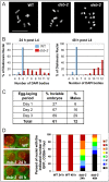
Figure 1
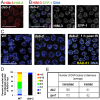
Figure 1. dsb-2 mutant worms exhibit a defect in CO/chiasma formation that worsens with age.
(A) Each panel shows the full complement of chromosomes in a single diakinesis-stage oocyte, stained by DAPI. Left: WT nucleus with six DAPI bodies corresponding to six pairs of homologs connected by chiasmata (bivalents). Middle: dsb-2(me96) nucleus with eight DAPI bodies (4 bivalents and 4 univalents), indicating that only four pairs of homologs have formed chiasmata. Right: dsb-2(me96) nucleus with 12 DAPI bodies (all univalents), indicating absence of chiasmata between all homolog pairs. Scale bar, 10 µm. (B) Graphs showing frequencies of diakinesis-stage nuclei with the indicated number of DAPI bodies in dsb-2(me96) and WT hermaphrodites fixed and stained at 1 day and 2 days post L4. (C) Table showing frequency of inviable embryos and frequency of males (among surviving progeny) from eggs laid by dsb-2(me96) rol-1(e91) hermaphrodites (where rol-1 is a marker that does not affect meiosis) during the indicated time interval after the L4 stage. Inviable embryos that do not hatch are indicative of autosomal mis-segregation, while male progeny indicate X-chromosome mis-segregation. For comparison, wild-type hermaphrodites produce less than 1% inviable embryos and approximately 0.2% males during their entire reproductive lives. (D) Left: images of GFP::COSA-1 foci in late pachytene nuclei of live anesthetized worms, with chromatin visualized by mCherry::H2B and plasma membranes marked by GFP::PH. Each WT nucleus has 6 GFP::COSA-1 foci, corresponding to the single CO site on each homolog pair; reduced numbers of GFP::COSA-1 foci in the dsb-2(me97) nuclei reflect reduced CO formation. Scale bar, 5 µm. Right: Graph showing frequencies of nuclei with indicated numbers of GFP::COSA-1 foci in late pachytene nuclei of worms examined at 24 or 48 h post L4, revealing worsening of the CO deficit with age in dsb-2(me97) mutant worms.
Figure 2
Figure 2. DSB-2 is required for meiotic DSB formation.
(A) Homolog pairing assayed by immunofluorescence of X-chromosome pairing center (X-PC) binding protein HIM-8 in dsb-2(me96) pachytene nuclei. A single HIM-8 focus is observed in each nucleus, indicating successful pairing at the X-PC. HIM-3 marks chromosome axes. Scale bar, 10 µm. (B) Synapsis assayed by immunofluorescence in dsb-2(me96) pachytene nuclei. Axis protein HIM-3 and SC central region protein SYP-1 co-localize in continuous stretches between chromosome pairs, indicating successful synapsis. Scale bar, 10 µm. (C) Immunolocalization of RAD-51 in early pachytene nuclei. RAD-51 foci mark DSBs and are greatly reduced or absent in untreated dsb-2(me96) nuclei compared to WT. Following irradiation (IR), RAD-51 foci are abundant in dsb-2(me96) nuclei, indicating that the dsb-2 mutant retains that capacity to load RAD-51 at induced DSBs. Scale bar, 10 µm. (D) Quantitation of reduced RAD-51 foci in the dsb-2(me96) mutant. RAD-51 foci were scored in 8 contiguous rows of pachytene nuclei from the region of the germ line where foci were most abundant in wild type (see Materials and Methods). (E) Table showing average numbers of DAPI bodies in diakinesis-stage oocytes in dsb-2(me96) and spo-11 mutant worms with and without irradiation, showing restoration of chiasma formation by IR-induced DSBs. Worms were exposed to 1 kRad of gamma-irradiation at 36 hours post L4, and irradiated and age-matched controls were fixed and stained with DAPI 18 hours post irradiation. As in Figure 1A and B, the number of DAPI bodies reflects success or failure of chiasma formation: 12 indicates lack of chiasmata for all homolog pairs, and 6 indicates successful chiasma for all homolog pairs. This assay tends to underestimate the incidence of achiasmate chromosomes, as some lie too close together to be resolved.
Figure 3
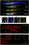
Figure 3. DSB-2 localizes to chromatin in early meiotic prophase nuclei, concurrent with DSB marker and SUN-1 Ser8-phosphorylation.
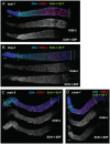
(A) Immunofluorescence image of a WT hermaphrodite gonad (from distal tip to end of pachytene) stained with DAPI and antibodies that recognize DSB-2 and SUN-1 S8P. In this and all subsequent figures, the distal tip (which contains mitotically cycling germ cells) is at the left, and meiotic progression is from left to right. DSB-2 first appears on chromatin at meiotic onset in transition zone nuclei (corresponding to the leptotene/zygotene stages of meiotic prophase) and disappears around mid-pachytene stage of meiotic prophase, with a few outlier nuclei retaining DSB-2 in later pachytene. Here and in subsequent figures, yellow lines demarcate the “DSB-2-positive” zone and a cyan line marks the end of the pachytene zone. Co-staining shows correlation between DSB-2-positive and SUN-1 S8P-positive meiotic nuclei. (Note: SUN-1 S8P is also present in a few pre-meiotic nuclei; [23]). A close-up of the large box is shown in Figure 5C. Scale bar, 15 µm. (B) Close-up of nuclei outlined by the small box in (A). DSB-2 localizes to a few bright patches/foci, as well as fainter stretches/foci along the entire chromatin (see also Fig. 5C). As nuclei reach mid-pachytene, the DSB-2 signal becomes fainter (narrow arrowhead), however in some nuclei signal gets brighter along most of chromatin (broad arrowhead). (C) Immunofluorescence image of a WT hermaphrodite gonad from entry into meiotic prophase to mid-to-late pachytene, stained with antibodies that recognize DSB-2 and RAD-51. RAD-51 foci (marking processed DSBs) appear in nuclei shortly after DSB-2 staining appears on chromatin upon meiotic entry, and the RAD-51 foci disappear shortly after DSB-2 is no longer present on chromatin in mid-pachytene nuclei. Inset shows that RAD-51 foci mostly do not co-localize with concentrated DSB-2. DSB-2-bright outlier nuclei in late pachytene contain high levels of RAD-51 foci. Scale bar, 15 µm. (D) Immunofluorescence image of the early mid-pachytene to late pachytene region of a WT hermaphrodite gonad expressing GFP::COSA-1 (strain AV630), stained with antibodies that recognize DSB-2 and GFP. COSA-1 foci marking designated CO sites appear in nuclei only after the removal of DSB-2 from chromatin; DSB-2-bright outlier nuclei in the late pachytene region lack COSA-1 foci, even when COSA-1 foci are present in neighboring nuclei. Close-ups are shown in insets. Scale bar, 15 µm.
Figure 4
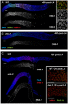
Figure 4. Relationship between DSB-2 and paralog DSB-1.
(A) Immunofluorescence image of a WT hermaphrodite gonad (from distal tip to end of pachytene), stained with DAPI and antibodies that recognize DSB-2 and DSB-1. DSB-2 and DSB-1 are detected in a highly correlated subset of germline nuclei (within a region spanning from meiotic prophase onset through mid-pachytene), although the relative intensities of the DSB-2 and DSB-1 signals vary during prophase progression (see text). Inset: close-up of the field of early pachytene nuclei outlined in (A) showing that although DSB-1 and DSB-2 are present in the same nuclei, the DSB-1 and DSB-2 signals on chromatin sometimes overlap but mostly do not match each other. Scale bar, 15 µm. (B, C) DSB-1 immunolocalization in the dsb-2(me96) mutant. Zoomed out images show that DSB-1 staining is fairly similar to wild-type control in a 12 h post-L4 dsb-2 mutant gonad (C), but DSB-1 staining is reduced (relative to wild-type) in the pachytene region of a 48 h post-L4 dsb-2 mutant gonad (B). Insets in C show fields of pachytene nuclei illustrating that RAD-51 foci are already reduced in the dsb-2 mutant at 12 h post L4. Scale bars, 15 µm.
Figure 5
Figure 5. DSB-2 and SUN-1 S8P persist when DSB formation is defective.
(A) and (B) Immunofluorescence images of gonads from the distal pre-meiotic region to end of pachytene, stained with DAPI and antibodies that recognize DSB-2 and SUN-1 S8P. The zone of DSB-2 and SUN-1 S8P-positive nuclei is extended in both spo-11 (A) and him-17 (B) mutants, which are defective in DSB formation. (C) Close-up images of fields of nuclei in early pachytene, as outlined in Figure 3A and (A), (B) above. WT as well as spo-11 nuclei show bright patches of DSB-2 staining, whereas him-17 nuclei do not. Scale bar, 15 µm.
Figure 6
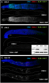
Figure 6. DSB-2 and SUN-1 S8P are coordinately regulated by common upstream regulator CHK-2.
Immunofluorescence images of gonads of indicated genotypes from the distal pre-meiotic region to end of pachytene, stained with DAPI and antibodies that recognize DSB-2 and SUN-1 S8P. Scale bar, 15 µm. (A) SUN-1 S8P is detected at the NE in meiotic prophase nuclei in the dsb-2 mutant germ line, indicating that although these features are coordinated during wild-type meiosis, acquisition of meiotic SUN-1 S8P does not depend on DSB-2. However, the SUN-1 S8P zone is extended in the dsb-2 mutant, indicating that the timing of its removal is affected. DSB-2 staining is absent from chromatin, indicating antibody specificity. (B) Main panel: Immunofluorescence images showing that localization of DSB-2 on chromatin and SUN-1 S8P staining at the NE are both severely reduced in the chk-2 mutant in the indicated meiotic region. Note: SUN-1 S8P signal remains present on some pre-meiotic nuclei and on late diakinesis oocytes in chk-2 mutants (data not shown; [23]). Inset: Western blot of whole-worm protein lysates from the indicated genotypes (60 worms per lane) stained with anti-DSB-2 antibodies. The arrow indicates the DSB-2 protein (32 kD), which is absent in the dsb-2 mutant but is still present in the chk-2 mutant; the asterisk indicates a non-specific band that serves as a loading control. (C) The presence of DSB-2 on chromatin and SUN-1 S8P at the NE are correlated in the him-19 mutant, in which only a small subset of nuclei are positive for these marks.
Figure 7
Figure 7. Quantitation of the DSB-2 positive zone in WT and meiotic mutants.
Bar graph showing the extent of the region of DSB-2 positive nuclei in germ lines of indicated genotypes. The presence/absence of DSB-2 signals was assessed in the portion of the germ line extending from the onset of DSB-2 staining to the end of the pachytene region. The extent of the DSB-2 positive zone was defined as the percentage of continuous rows of nuclei in which all or most nuclei exhibited DSB-2 staining out of total rows of nuclei in the scored region. Representative germ lines were imaged and scored: 5 for WT and 3 for each of the meiotic mutants. Error bars show standard deviation.
Figure 8
Figure 8. DSB-2 and SUN-1 S8P persist in mutants defective for DSB repair.
Immunofluorescence images of gonads of indicated genotypes from the distal pre-meiotic region to end of pachytene, stained with DAPI and antibodies that recognize DSB-2 and SUN-1 S8P. The zone of DSB-2 and SUN-1 S8P-positive nuclei is extended in all three mutants depicted: (A) rad-50, which is defective in both DSB formation and DSB repair; (B) and (C) rad-51 and rad-54, which are defective in DSB repair. In the rad-50 mutant germ line, pachytene nuclei have variable staining intensities, with some bright DSB-2 and/or SUN-1 S8P-positive nuclei scattered over the entire pachytene zone; this likely reflects the fact that although the rad-50 mutant lacks SPO-11-dependent DSBs, many nuclei enter meiotic prophase with existing DNA damage resulting from failure to repair lesions arising during DNA replication . DSB-2 staining persists until the end of the pachytene region of the rad-51 and rad-54 mutant gonads, which are also shorter than the gonads of wild-type controls. Scale bar, 15 µm.
Figure 9
Figure 9. DSB-2 and SUN-1 S8P persist when CO formation is impaired.
Immunofluorescence images of gonads of indicated genotypes from the distal pre-meiotic region to end of pachytene, stained with DAPI and antibodies that recognize DSB-2 and SUN-1 S8P. The zone of DSB-2 and SUN-1 S8P-positive nuclei is extended in: (A) the syp-1 mutant, which is defective for SC formation and for formation of interhomolog COs; and (B, C and D) the zhp-3, msh-5 and cosa-1 mutants, respectively, which are proficient for synapsis and DNA repair but are defective in conversion of DSBs to COs. Scale bar, 15 µm.
Figure 10
Figure 10. DSB-2 and SUN-1 S8P persistence requires axis proteins HTP-1 and HTP-3.
(A and B) Immunofluorescence images of gonads of indicated genotypes from the distal pre-meiotic region to end of pachytene, stained with DAPI and antibodies that recognize DSB-2 and SUN-1 S8P. The zone of DSB-2 and SUN-1 S8P-positive nuclei is not extended in the htp-1 and htp-3 mutants, which lack major components of the meiotic chromosome axes, despite the fact that these mutants are impaired in formation of interhomolog COs.
Figure 11
Figure 11. DSB-2 marked nuclei require RAD-50 for formation of RAD-51 foci after irradiation.
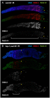
Immunofluorescence images of rad-50 (A) and htp-1; rad-50 (B) mutant gonads from the distal pre-meiotic region to end of pachytene, stained with DAPI and antibodies that recognize DSB-2 and RAD-51. Worms were fixed and stained 1 hour after exposure to 5 kRad of gamma-irradiation. A reciprocal relationship is observed between DSB-2 and RAD-51 immunolocalization: in nuclei where DSB-2 signal is detected on chromatin, formation of irradiation-induced RAD-51 foci is inhibited, and in nuclei where IR-induced RAD-51 foci are present, DSB-2 is absent. The zone of DSB-2 staining/RAD-51 inhibition is indicated by brackets. (Occasional bright RAD-51 foci in the “inhibited” zone are thought to represent pre-existing DNA damage acquired during mitotic cell cycles in mutants lacking RAD-50, as they are both irradiation- and SPO11-independent .) Arrowheads point to examples of nuclei that retain DSB-2 staining/RAD-51 inhibition in a region of the germ line where their neighbors do not. Scale bar, 15 µm. While the zone of DSB-2 staining/RAD-51 inhibition in the irradiated rad-50 single mutant extends from meiotic prophase entry to late pachytene, the zone of DSB-2 staining/RAD-51 inhibition is limited to a smaller domain from meiotic entry to the early pachytene region in the irradiated htp-1; rad-50 double mutant.
Figure 12
Figure 12. Model for regulatory network coordinating DSB competence with other aspects of meiotic progression.
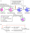
(A) Diagram depicting progression of germ cells through meiotic prophase. DSB-2 localization to chromatin (magenta) and SUN-1 S8P (green) at the nuclear envelope (NE) indicate a cellular state in which chromatin is permissive for DSB formation by SPO-11. Once a germ cell senses that sufficient CO-eligible inter-homolog (IH) intermediates have been formed to guarantee one CO per chromosome pair, the cell responds by shutting down both DSB formation and several specialized features of the meiotic mode of DNA repair (access to the homolog as a repair partner, dependence on RAD-50 for RAD-51 loading). This coordinate transition corresponds to removal of DSB-2 from chromatin and loss of SUN-1 S8P, and enables progression to CO designation and maturation of CO sites. In some nuclei, persistent DNA damage and/or lack of CO-eligible IH intermediates can lead to increased DSB-2 on chromatin (purple) and persistence of SUN-1 S8P at the NE, features that may mark germ cells destined for apoptosis. (B) The CHK-2 protein kinase acts as a common upstream regulator of both DSB-2 localization on chromatin and SUN-1 S8P at the NE, which function in parallel to promote competence for DSB formation (DSB-2) and homologous chromosome synapsis (SUN-1 S8P). Both of these processes are essential for formation of inter-homolog COs. The emergence of sufficient CO-eligible repair intermediates leads to negative feedback regulation by which DSB-2 and SUN-1 S8P are removed, shutting down DSB formation and leading to a change in nucleus state and properties. This coordinated negative feedback regulation may occur through common regulator CHK-2.

References

    1. Keeney S (2001) Mechanism and control of meiotic recombination initiation. Curr Top Dev Biol 52: 1–53. - PubMed
    1. Page SL, Hawley RS (2003) Chromosome choreography: the meiotic ballet. Science 301: 785–789. - PubMed
    1. Keeney S (2008) Spo11 and the Formation of DNA Double-Strand Breaks in Meiosis. Genome Dyn Stab 2: 81–123. - PMC - PubMed
    1. Kumar R, Bourbon HM, de Massy B (2010) Functional conservation of Mei4 for meiotic DNA double-strand break formation from yeasts to mice. Genes Dev 24: 1266–1280. - PMC - PubMed
    1. Lake CM, Nielsen RJ, Hawley RS (2011) The Drosophila zinc finger protein trade embargo is required for double strand break formation in meiosis. PLoS Genet 7: e1002005. - PMC - PubMed

Publication types

MeSH terms

Substances