. 2013;9(8):e1003715.
doi: 10.1371/journal.pgen.1003715. Epub 2013 Aug 8.

Global analysis of fission yeast mating genes reveals new autophagy factors

Affiliations

Global analysis of fission yeast mating genes reveals new autophagy factors

Ling-Ling Sun et al. PLoS Genet. 2013.

Abstract

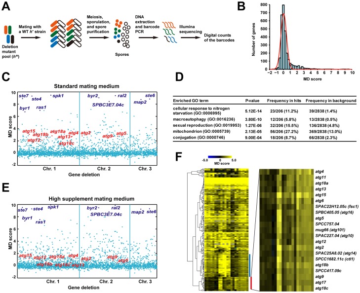
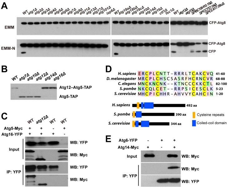
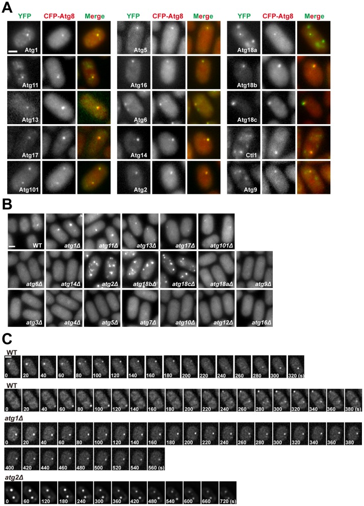
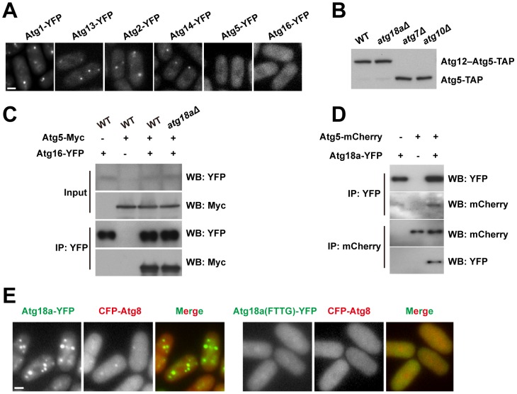
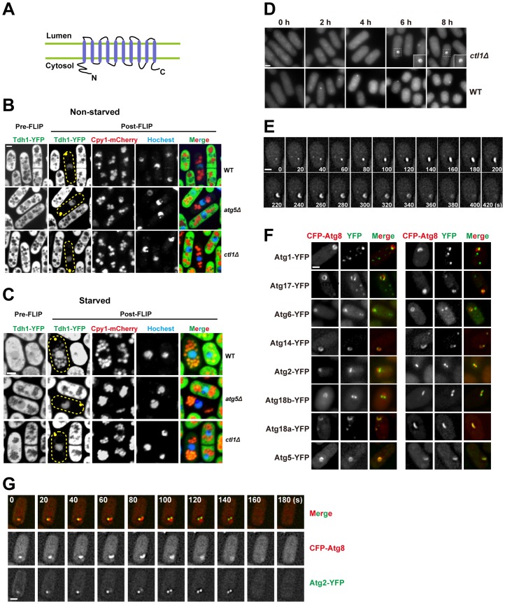
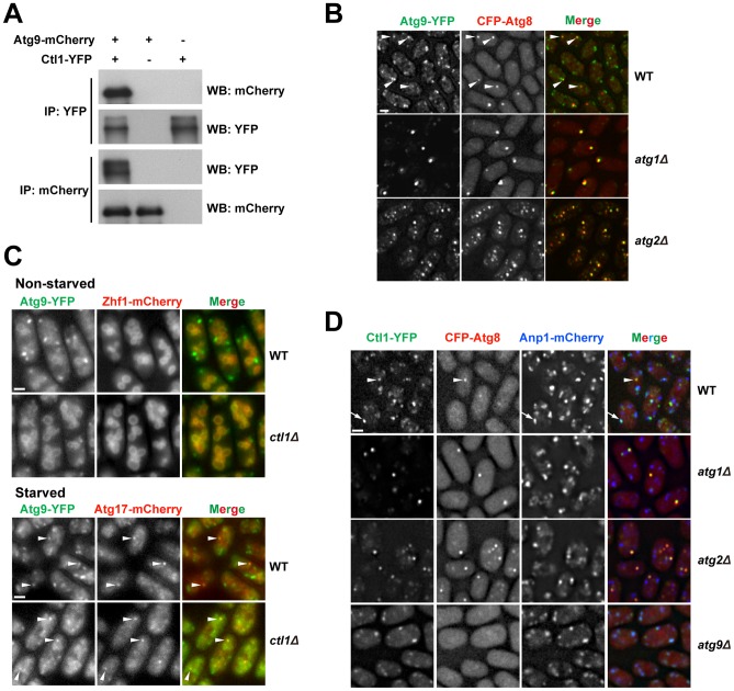
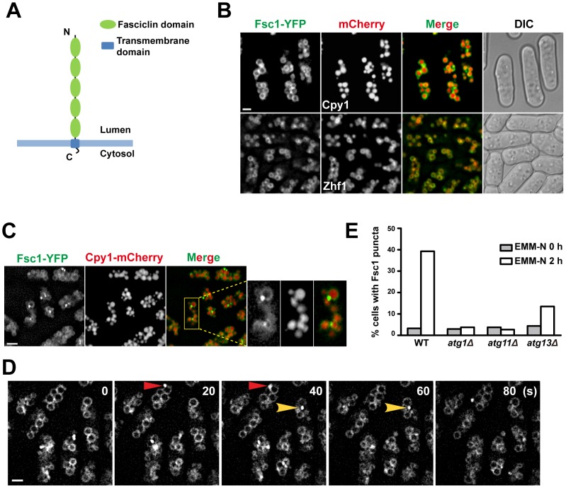
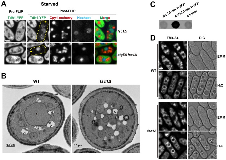
Macroautophagy (autophagy) is crucial for cell survival during starvation and plays important roles in animal development and human diseases. Molecular understanding of autophagy has mainly come from the budding yeast Saccharomyces cerevisiae, and it remains unclear to what extent the mechanisms are the same in other organisms. Here, through screening the mating phenotype of a genome-wide deletion collection of the fission yeast Schizosaccharomyces pombe, we obtained a comprehensive catalog of autophagy genes in this highly tractable organism, including genes encoding three heretofore unidentified core Atg proteins, Atg10, Atg14, and Atg16, and two novel factors, Ctl1 and Fsc1. We systematically examined the subcellular localization of fission yeast autophagy factors for the first time and characterized the phenotypes of their mutants, thereby uncovering both similarities and differences between the two yeasts. Unlike budding yeast, all three Atg18/WIPI proteins in fission yeast are essential for autophagy, and we found that they play different roles, with Atg18a uniquely required for the targeting of the Atg12-Atg5·Atg16 complex. Our investigation of the two novel factors revealed unforeseen autophagy mechanisms. The choline transporter-like protein Ctl1 interacts with Atg9 and is required for autophagosome formation. The fasciclin domain protein Fsc1 localizes to the vacuole membrane and is required for autophagosome-vacuole fusion but not other vacuolar fusion events. Our study sheds new light on the evolutionary diversity of the autophagy machinery and establishes the fission yeast as a useful model for dissecting the mechanisms of autophagy.


Conflict of interest statement

The authors have declared that no competing interests exist.

Figures

Figure 1
Figure 1. Barcode sequencing-based screens of mating phenotype.
(A) Schematic of the screening procedure. (B) A histogram of the mating defect (MD) scores from the screen 0428_YES_SPA-45s conducted under standard mating conditions. The red line represents a fitted normal distribution. (C) A scatter plot of the MD scores from the screen 0428_YES_SPA-45s. The genes are ordered according to their chromosomal positions. The 10 genes with the highest average MD scores under standard conditions are highlighted in dark blue. The 10 genes known to be required for starvation-induced autophagy are highlighted in red. (D) Gene Ontology (GO) term enrichment analysis of the screen hits obtained under standard conditions. (E) A scatter plot of the MD scores from the screen 0428_YES_SPA-200s. Genes are highlighted as in C. (F) Hierarchical clustering analysis of the MD scores from the 22 screens. For a detailed view of the heat map, see Figure S2. Blue bar denotes the cluster enriched for mitochondrial protein-coding genes. Red bar denotes the autophagy gene cluster, whose close-up view is shown at right.
Figure 2
Figure 2. CFP-Atg8 processing defect of autophagy mutants and the identification of Atg10, Atg16, and Atg14.
(A) CFP-Atg8 processing assay. Cells were collected before and 8 h after shifting to a nitrogen-free medium (EMM-N). (B) The conjugation of Atg12 to Atg5 requires the atg10 gene. (C) Atg5-Myc was co-immunoprecipitated with Atg16-YFP in both wild-type and atg12Δ cells. Input, 1%; IP, 20%. (D) The “cysteine repeats” region and the domain organization of Atg14 proteins. Coiled-coil domains are predicted as in Figure S4. (E) Atg14-Myc was co-immunoprecipitated with Atg6-YFP. Input, 1%; IP, 20%.
Figure 3
Figure 3. Subcellular localization of fission yeast autophagy factors.
(A) Fifteen Atg proteins colocalized with CFP-Atg8 at cytoplasmic puncta induced by starvation. Images were acquired 2 h after starvation. (B) The distribution of CFP-Atg8 in atg mutants. Images were acquired 3 h after starvation. (C) Time-lapse analysis of CFP-Atg8 puncta in wild type, atg1Δ, and atg2Δ cells. Bars, 3 µm.
Figure 4
Figure 4. Atg18a is required for the PAS targeting of the Atg12–Atg5·Atg16 complex.
(A) atg18aΔ abolished the starvation-induced puncta formation by Atg5 and Atg16. (B) Atg12–Atg5 conjugation is normal in atg18aΔ cells. (C) The interaction between Atg5 and Atg16 is intact in atg18aΔ cells. Input, 1%; IP, 20%. (D) Atg5 and Atg18a co-immunoprecipitated with each other. (E) Mutating the FRRG motif in Atg18a abolished its own puncta and the Atg8 puncta in starved cells. Bars, 3 µm.
Figure 5
Figure 5. Ctl1 is required for autophagic transport and normal PAS organization.
(A) The predicted membrane topology of Ctl1. (B,C) A fluorescence loss in photobleaching (FLIP) assay revealed the immobilized and non-diffusible pools of Tdh1-YFP. Yellow dots mark the sites of photobleaching. (B) In non-starved cells, only nuclear YFP signal remained in post-FLIP images. (C) In starved cells, vacuolar YFP signal was observed in post-FLIP images of wild-type, but not atg5Δ or ctl1Δ cells. (D) Ring-shaped and C-shaped structures labeled by CFP-Atg8 appeared in ctl1Δ cells after prolonged starvation. (E) Time-lapse images of a ctl1Δ cell. (F) The localization patterns of other Atg proteins in ctl1Δ cells containing CFP-Atg8-labeled structures. (G) Time-lapse images of a ctl1Δ cell expressing both CFP-Atg8 and YFP-Atg2. Bars, 3 µm.
Figure 6
Figure 6. Ctl1 and Atg9 interact with each other and influence each other's localization.
(A) Atg9 and Ctl1 co-immunoprecipitated with each other. (B) Localization patterns of Atg9 and Atg8 in starved cells. Arrowheads point to the puncta where Atg9 and Atg8 colocalized in the wild-type cells. (C) ctl1Δ altered the localization pattern of Atg9 in non-starved cells. Zhf1 is a vacuole membrane marker , and Atg17 is a PAS marker. Arrowheads point to puncta where Atg9 and Atg17 colocalized. (D) Localization patterns of Ctl1 in starved cells. Atg8 and Anp1 are PAS and Golgi markers, respectively. The arrowhead points to a punctum where Ctl1 and Atg8 colocalized. The arrow points to a punctum where Ctl1 and Anp1 colocalized. Bars, 3 µm.
Figure 7
Figure 7. Fsc1 localizes to the vacuole membrane and forms starvation-induced puncta.
(A) The predicted membrane topology and domain organization of Fsc1. (B) Localization of Fsc1 in non-starved cells. Cpy1 and Zhf1 are vacuole lumen and vacuole membrane markers, respectively. Bar, 3 µm. (C) Localization of Fsc1 in starved cells. Bar, 6 µm. (D) Time-lapse images of Fsc1 puncta induced by starvation. Bar, 3 µm. (E) Fsc1 puncta induced by starvation are dependent on Atg1, Atg11, and partially dependent on Atg13. Two hundred cells were examined for each data point.
Figure 8
Figure 8. Fsc1 is required for autophagosome-vacuole fusion.
(A) In starved fsc1Δ cells, Tdh1-YFP entered closed cytoplasmic membrane structures, which are not vacuoles. These structures are dependent on Atg5, thus are likely autophagosomes. Bar, 3 µm. (B) TEM analysis of starved wild-type and fsc1Δ cells. N, nucleus; M, mitochondrion; V, vacuole; AP, autophagosome. (C) Unlike the mutant lacking a general vacuolar fusion factor Aut12, fsc1Δ cells did not secret Cpy1-YFP, which was detected by a colony blot assay with an antibody recognizing YFP. The control is a strain not expressing Cpy1-YFP. (D) fsc1Δ did not affect homotypic vacuole fusion occurring when cells were shifted from EMM medium to water. Vacuoles were stained with the vital dye FM4-64. Bar, 3 µm.

References

    1. Takeshige K, Baba M, Tsuboi S, Noda T, Ohsumi Y (1992) Autophagy in yeast demonstrated with proteinase-deficient mutants and conditions for its induction. J Cell Biol 119: 301–311. - PMC - PubMed
    1. Tsukada M, Ohsumi Y (1993) Isolation and characterization of autophagy-defective mutants of Saccharomyces cerevisiae. FEBS Lett 333: 169–174. - PubMed
    1. Kohda TA, Tanaka K, Konomi M, Sato M, Osumi M, et al. (2007) Fission yeast autophagy induced by nitrogen starvation generates a nitrogen source that drives adaptation processes. Genes Cells 12: 155–170 doi:10.1111/j.1365-2443.2007.01041.x - DOI - PubMed
    1. Levine B, Kroemer G (2008) Autophagy in the pathogenesis of disease. Cell 132: 27–42 doi:10.1016/j.cell.2007.12.018 - DOI - PMC - PubMed
    1. Mizushima N, Komatsu M (2011) Autophagy: renovation of cells and tissues. Cell 147: 728–741 doi:10.1016/j.cell.2011.10.026 - DOI - PubMed

Publication types