. 2013 Aug 7;8(8):e70795.
doi: 10.1371/journal.pone.0070795. eCollection 2013.

Trypsin potentiates human fibrocyte differentiation

Affiliations

Trypsin potentiates human fibrocyte differentiation

Michael J V White et al. PLoS One. .

Abstract

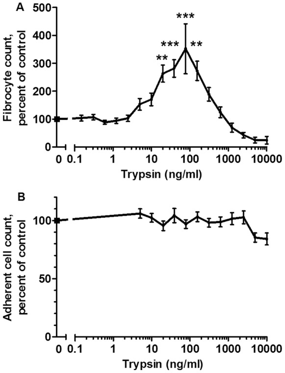
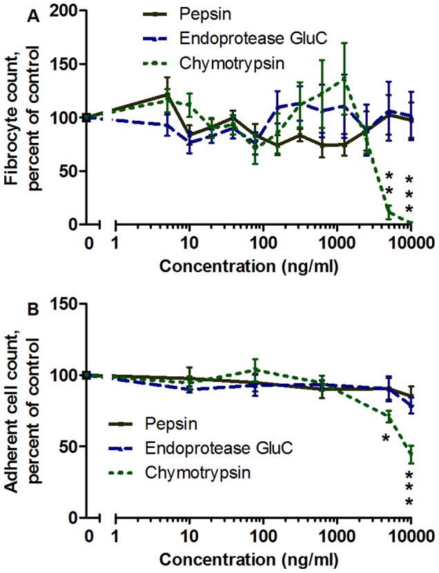
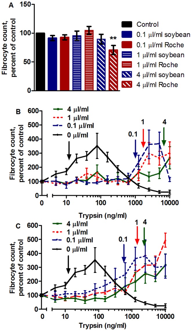
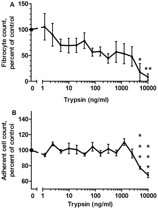
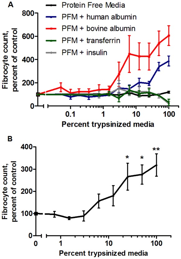
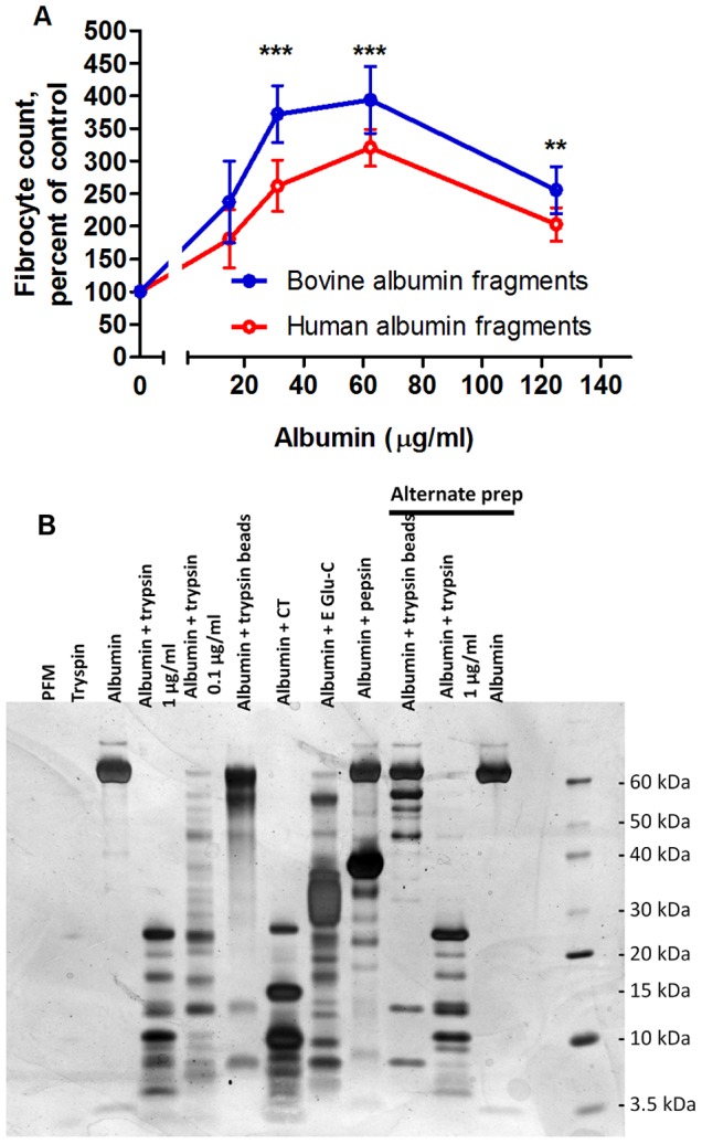
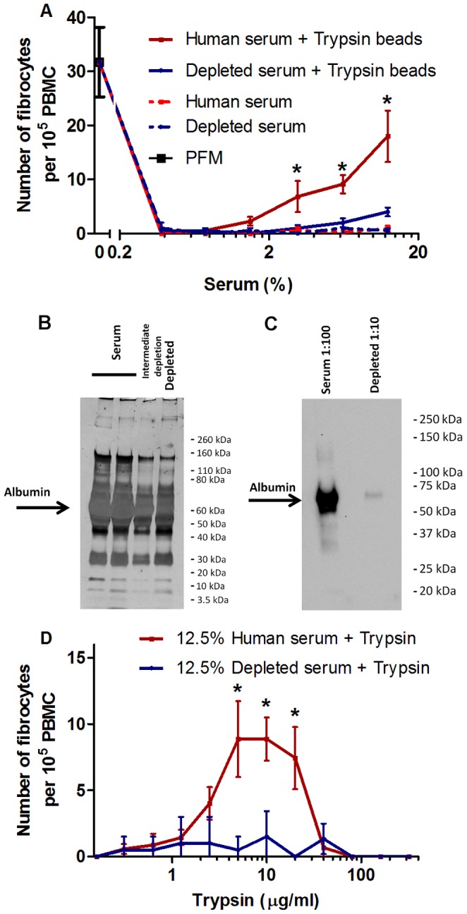
Trypsin-containing topical treatments can be used to speed wound healing, although the mechanism of action is unknown. To help form granulation tissue and heal wounds, monocytes leave the circulation, enter the wound tissue, and differentiate into fibroblast-like cells called fibrocytes. We find that 20 to 200 ng/ml trypsin (concentrations similar to those used in wound dressings) potentiates the differentiation of human monocytes to fibrocytes in cell culture. Adding trypsin inhibitors increases the amount of trypsin needed to potentiate fibrocyte differentiation, suggesting that the potentiating effect is dependent on trypsin proteolytic activity. Proteases with other site specificities such as pepsin, endoprotease GluC, and chymotrypsin do not potentiate fibrocyte differentiation. This potentiation requires the presence of albumin in the culture medium, and tryptic fragments of human or bovine albumin also potentiate fibrocyte differentiation. These results suggest that topical trypsin speeds wound healing by generating tryptic fragments of albumin, which in turn potentiate fibrocyte differentiation.


Conflict of interest statement

Competing Interests: The authors have declared that no competing interests exist.

Figures

Figure 1
Figure 1. Trypsin potentiates fibrocyte differentiation.
(A) PBMC were cultured in serum free media in the presence of the indicated concentrations of trypsin for 5 days, after which the PBMC were air-dried, stained, counted for fibrocyte differentiation, and normalized for each donor to the no-trypsin control. The no-trpysin controls developed 41.8±5.4 fibrocytes per 105 PBMC. (B) The same PBMC populations were then counted for the total number of PBMC adhered to the plate following fixing and staining and normalized for each donor to the no-trypsin control. There were no significant differences in the numbers of adhered PBMC following fixing and staining. Values in A and B are mean ± SEM, n = 9. **indicates p<.01 and ***indicates p<.001 compared to the no-trypsin control by 1-way ANOVA, Dunnett’s test.
Figure 2
Figure 2. Chymotrypsin, pepsin, and endoproteinase GluC do not potentiate fibrocyte differentiation.
(A) PBMC were cultured with the indicated concentrations of protease for 5 days, and fibrocytes were counted as in figure 1. (B) The same PBMC populations were then counted for the total number of PBMC adhered to the plate following fixing and staining, and normalized to the no-protease control. At high concentrations, chymotrypsin significantly lowered the numbers of fibrocytes and adhered PBMC. Values are mean ± SEM, n = 9 for pepsin and endoproteinase GluC, and n = 7 for chymotrypsin. *indicates p<.05, **indicates p<.01, and ***indicates p<.001 compared to the no-protease control by 1-way ANOVA, Dunnett’s test.
Figure 3
Figure 3. Trypsin inhibitors increase the amount of trypsin needed to potentiate fibrocyte potentiation.
Trypsin inhibitors were added to PBMC cultures at the beginning of the 5-day incubation period at the indicated concentrations, with and without trypsin present. PBMC were then air dried, stained, counted for fibrocyte differentiation, and normalized to the no-trypsin control. (A) Trypsin inhibitors did not significantly affect fibrocyte differentiation with the exception of 4 µl/ml Roche inhibitor, which decreased fibrocyte formation. **indicates p<.01 compared to the inhibitor-free control. (B) Soybean trypsin inhibitor inhibited trypsin-induced fibrocyte potentiation. No-inhibitor data is the same as figure 1A. Fibrocyte counts were normalized to the inhibitor-containing trypsin-free control. Arrows indicate the lowest trypsin concentrations that doubled the fibrocyte number. Compared to the no-trypsin control, there was a significant increase in the number of fibrocytes with p<.05 for 4 µl/ml inhibitor for 10,000 ng/ml and at 1 µl/ml inhibitor for 5,000 ng/ml, and p<.001 at 1 µl/ml inhibitor for 2,500 and 10,000 ng/ml and at 0.1 µl/ml inhibitor for 2500 and 5,000 ng/ml (1-way ANOVA, Dunnett’s test). (C) Roche complete protease inhibitor cocktail inhibitor in the cell culture medium also inhibited trypsin-induced fibrocyte potentiation. No-inhibitor data is the same as figure 1A. Arrows indicate the lowest trypsin concentrations that doubled the fibrocyte number. Compared to the no-trypsin control, there was a significant increase in the number of fibrocytes with p<.05 at 4 µl/ml inhibitor for 1250 ng/ml and 0.1 µl/ml inhibitor for 1250 and 2500 ng/ml, p<.01 at 1 µl/ml inhibitor for 10,000 ng/ml and at 0.1 µl/ml inhibitor for 10,000 ng/ml, and p<.001 at 4 µl/ml inhibitor for 2500, 5000, and 10,000 ng/ml (1-way ANOVA, Dunnett’s test). Values are mean ± SEM, n = 7.
Figure 4
Figure 4. Trypsin does not potentiate fibrocyte differentiation in medium lacking protein supplements.
(A) PBMC were cultured with the indicated concentrations of trypsin for 5 days in protein-free medium, after which the PBMC were air-dried, stained, counted for fibrocyte differentiation, and normalized to the no-trypsin control. The no-trypsin controls developed 39±7.2 fibrocytes per 105 PBMC. (B) The same PBMC populations were then counted for the total number of PBMC adhered to the plate following fixing and staining. Values are mean ± SEM, n = 7. *indicates p<.05, **indicates p<.01, and ***indicates p<.001 compared to the no-trypsin control by 1-way ANOVA, Dunnett’s test.
Figure 5
Figure 5. Serum-free medium containing albumin potentiates fibrocyte differentiation after temporary mixing with trypsin-coated agarose beads.
TPCK-treated trypsin-coated agarose beads were used to digest protein-free culture media (PFM) and PFM containing bovine albumin, human albumin, transferrin, or insulin, after which the beads were removed. Digested media and controls were mixed with SFM at the indicated percentages and added to PBMC at the beginning of a 5 day incubation, after which the PBMC were air-dried, stained, counted for fibrocyte differentiation, and normalized to the control media containing the same amount of undigested protein. (A) After removal of the beads, only medium containing albumin fragments potentiated fibrocyte differentiation. Values are mean ± SEM, n = 3 for protein-free medium (PFM), insulin, and transferrin, n = 4 for bovine albumin, and n = 7 for human albumin. Compared to the human albumin control, digested human albumin significantly increased the number of fibrocytes with p<.05 at 62 µg/ml, p<.01 at 250 µg/ml, and p<.001 at 500 µg/ml. Compared to the bovine albumin control, digested bovine albumin increased the number of fibrocytes with p<.05 at 31 µg/ml, p<.01 at 250 µg/ml, and p<.001 at 500 µg/ml (1-way ANOVA, Dunnett’s test). (B) Human albumin-containing media potentiated fibrocyte differentiation when digested by non-TPCK treated trypsin-coated agarose beads. Values are mean ± SEM, n = 3. *indicates p<.05 and **indicates p<.01 compared to the human albumin control by 1-way ANOVA, Dunnett’s test.
Figure 6
Figure 6. Albumin potentiates fibrocyte differentiation after temporary mixing with trypsin-coated agarose beads.
TPCK-treated trypsin-coated agarose beads were used to digest bovine or human albumin. After the beads were removed, the digested albumin was added to PBMC cultures in SFM at the indicated concentrations for 5 days, after which the PBMC were air-dried, stained, counted for fibrocyte differentiation, and normalized to the bovine or human albumin controls. Values are mean ± SEM, n = 4, **indicates p<.01 and ***indicates p<.001 for both bovine and human albumin compared to the no-albumin control (1-way ANOVA, Dunnett’s test). (B) 250 µl of protein-free media supplemented with human albumin was digested for two hours with gentle rotation at 37°C with 1 µg/ml protease, 0.1 µg/ml protease, or with 12.5 µg/ml TPCK-treated trypsin-coated agarose beads. Samples were then run on a 7.5% SDS-PAGE gel and silver stained. PFM indicates protein free medium, CT indicates chymtotrypsin, and Glu-C indicates endoprotease Glu-C. Alternative prep indicates samples from a repeat of this experiment that happened to be included in this gel. This gel is a digestion of human albumin, and is representative of similar gels of digestions of medium containing bovine albumin or ITS-3.
Figure 7
Figure 7. Collagen production is increased in PBMC exposed to trypsinized albumin.
(A) TPCK-treated trypsin-coated agarose beads were used to digest protein-free media and media containing human albumin. After the beads were removed, the media was added to PBMC cultures in serum-free medium at a 33% concentration. Cells were exposed to trypsinized human albumin for 5 days, then resuspended, stained with anti-collagen and alexa-fluor secondary antibodies, and measured for fluorescence by flow cytometry. Values are mean ± SEM, n = 9, *indicates p<.05 by two tailed Mann- Whitney’s t-test. (B) Representative fluorescence overlay for collagen staining. The cells in the left plot were stained with isotype control primary antibodies, while the cells in the right plot were stained with anti-collagen primary antibodies. Red indicates trypsin-treated cells, while black indicates no trypsin treatment of the cells.
Figure 8
Figure 8. Trypsinized human serum containing albumin potentiates fibrocyte differentiation.
(A) TPCK-treated trypsin-coated agarose beads were used to digest human serum and albumin-depleted human serum, which were then added to PBMC in protein-free media at the indicated concentrations for 5 days, after which the cells were air-dried, stained, counted for fibrocyte differentiation. Values are mean ± SEM, n = 7 for serum, n = 4 for depleted serum. Compared to the trypsinized depleted serum, trypsinized serum significantly increased the number of fibrocytes: *indicates p<.05 by a two-tailed Mann-Whitney’s t-test. Compared to depleted serum, trypsinized depleted serum did not significantly increase fibrocyte number by a two-tailed Mann-Whitney’s t-test. (B) Albumin-depleted serum shows less albumin, but a similar protein pattern to human serum, when both are run at 1∶10 dilutions on a silver stained 4–20% SDS-PAGE gel. (C) A Western blot of serum and albumin-depleted serum stained with anti human-albumin antibodies. (D) Trypsin was added to PBMC in a 12.5% serum containing medium, and a 12.5% albumin-depleted serum containing medium, at the indicated concentrations for 5 days, after which the cells were air-dried, stained, counted for fibrocyte differentiation. Values are mean ± SEM, n = 7 for serum, n = 4 for depleted serum. Compared to the trypsinized depleted serum, trypsinized serum significantly increased the number of fibrocytes: *indicates p<.05 by a two-tailed Mann-Whitney’s t-test.

Similar articles

Cited by

References

    1. Drew P, Posnett J, Rusling L (2007) The cost of wound care for a local population in England. Int Wound J 4: 149–155. - PMC - PubMed
    1. Hurd T, Posnett J (2009) Point prevalence of wounds in a sample of acute hospitals in Canada. Int Wound J 6: 287–293. - PMC - PubMed
    1. Vowden KR, Vowden P (2009) The prevalence, management and outcome for acute wounds identified in a wound care survey within one English health care district. J Tissue Viability 18: 7–12. - PubMed
    1. Vowden KR, Vowden P (2009) A survey of wound care provision within one English health care district. J Tissue Viability 18: 2–6. - PubMed
    1. Srinivasaiah N, Dugdall H, Barrett S, Drew PJ (2007) A point prevalence survey of wounds in north-east England. J Wound Care 16: 413–416, 418–419. - PubMed

Publication types

LinkOut - more resources