. 2013 Aug 9;8(8):e70849.
doi: 10.1371/journal.pone.0070849. eCollection 2013.

Cannabinoid receptor CB2 modulates axon guidance

Affiliations

Cannabinoid receptor CB2 modulates axon guidance

Gabriel Duff et al. PLoS One. .

Abstract

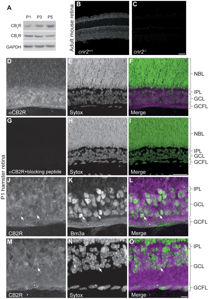
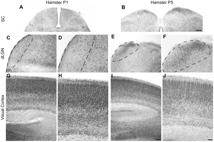
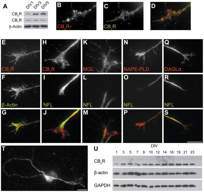
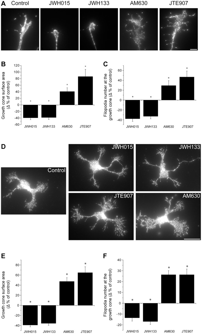
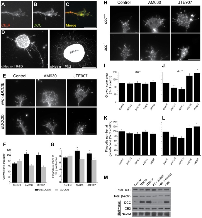
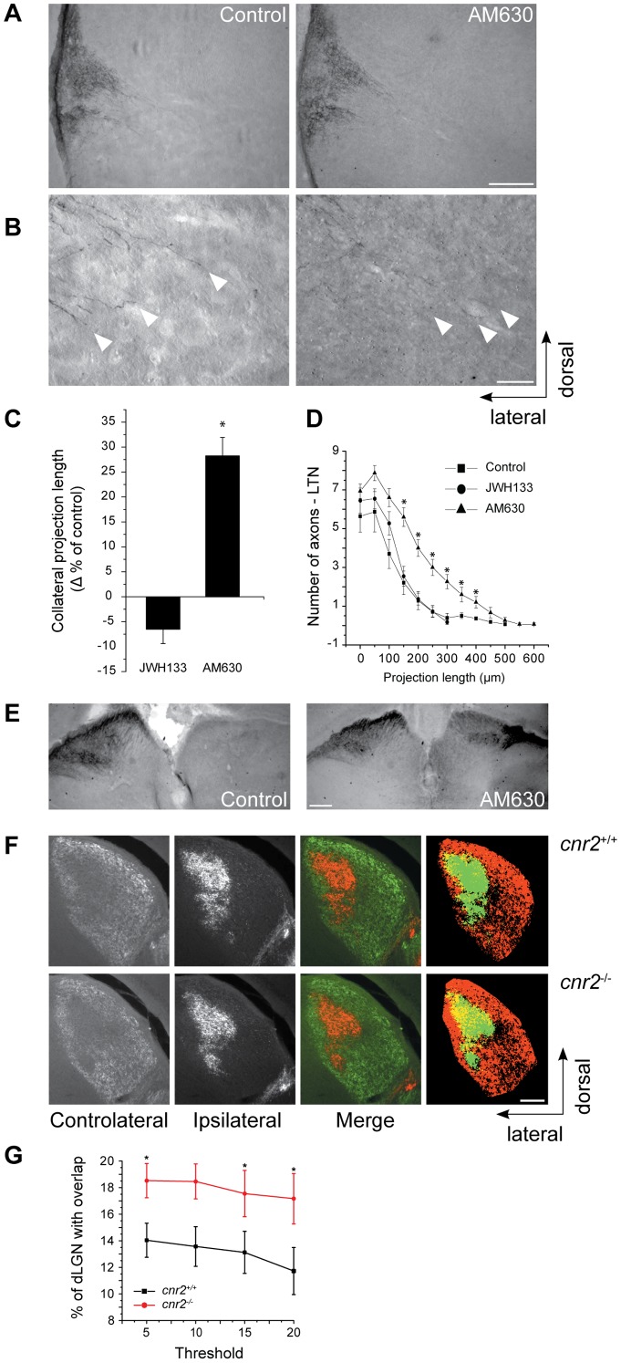
Navigation of retinal projections towards their targets is regulated by guidance molecules and growth cone transduction mechanisms. Here, we present in vitro and in vivo evidences that the cannabinoid receptor 2 (CB2R) is expressed along the retino-thalamic pathway and exerts a modulatory action on axon guidance. These effects are specific to CB2R since no changes were observed in mice where the gene coding for this receptor was altered (cnr2 (-/-)). The CB2R induced morphological changes observed at the growth cone are PKA dependent and require the presence of the netrin-1 receptor, Deleted in Colorectal Cancer. Interfering with endogenous CB2R signalling using pharmacological agents increased retinal axon length and induced aberrant projections. Additionally, cnr2 (-/-) mice showed abnormal eye-specific segregation of retinal projections in the dorsal lateral geniculate nucleus (dLGN) indicating CB2R's implication in retinothalamic development. Overall, this study demonstrates that the contribution of endocannabinoids to brain development is not solely mediated by CB1R, but also involves CB2R.


Conflict of interest statement

Competing Interests: The authors have declared that no competing interests exist.

Figures

Figure 1
Figure 1. Spatio-Temporal Expression of the CB2R in the Retina during Postnatal Retinal Projection Development in vivo.
(A) Western blot analysis of CB2R and CB1R expression during retinal postnatal development in the hamster. Photomicrographs illustrating the specificity of the CB2R antibody in the adult mouse retina (B, C) and the P1 hamster retina (D–I). (D–O), Photomicrographs of hamster retinal cross-sections showing CB2R (magenta) during early postnatal development (at postnatal day 1, P1). Sytox (green) was used to stain cell nuclei. Brn3a was used to label retinal ganglion cells (green). In panels (J–O), some CB2R positive retinal ganglion cell somas and fibers are indicated using arrows and asterisks respectively. NBL, Neuroblast layer; IPL, Inner plexiform layer; GCL, Ganglion cell layer; GCFL, Ganglion cell fiber layer. Specificity of the CB2R antibody is confimed using cnr2 +/+ and cnr2 /− adult mouse retina. Scale bars: 50 µm (B, C); 25 µm (D–I); 10 µm (J–O).
Figure 2
Figure 2. Expression of CB2R in the Superior Colliculus, dorsal Lateral Geniculate Nucleus, and Visual Cortex during Development in the Hamster.
Photomicrographs of coronal sections illustrating CB2R expression at P1 (A, C, D, G, H) and P5 (B, E, F, I, J) in the superior colliculus (SC) (A, B), the dorsal lateral geniculate nucleus (dLGN) (C–F), and the visual cortex (G–J). In panel C–F, dLGN has been outlined for better visualization. Scale bars: 200 µm (A, B); 100 µm (C, E, G, I); 50 µm (D, F, H, J).
Figure 3
Figure 3. Expression of CB2R, CB1R, MGL, NAPE-PLD, and DAGLα in vitro.
(A) Western blot shows temporal protein expression of CB2R and CB1R in cultured retinal ganglion cells. DIV1 retinal explants growth cones labeled with primary antibodies directed against CB2R (B, E, and H), CB1R (C), MGL (K), NAPE-PLD (N), and DAGLα (Q). Growth cones were also labeled for β-actin (F), Neurofilament-L (I, L, O, and R). Merged images are presented in D, G, J, M, P, and S. (T) Photomicrograph of a DIV2 primary cortical neuron immunolabeled for CB2R. Primary neurons were cultured for various numbers of days in vitro and cell extracts were equalized for total protein content. (U) Western blot showing temporal protein expression of CB2R, β-actin and GAPDH. Scale bars: 5 µm (B–S), 15 µm (T).
Figure 4
Figure 4. CB2R Reorganizes the Growth Cone Morphology.
(A) Retinal explants and (D) dissociated neurons were grown for 1 and 2 days in vitro, respectively. Growth cones were exposed for 1 hour to 300 nM JWH015, 300 nM JWH133, 300 nM AM630, or 300 nM JTE907. Following treatment, retinal explants and neurons were fixed and immunolabeled for L1 and GAP-43, respectively. Addition of CB2R inverse agonists (AM630, JTE907) increased growth cone surface area (B and E) and filopodia number (C and F) while the opposite effects was observed following CB2R agonists (JWH015 and JWH133) treatment (mean ± SEM; n = 374 to 714 per condition). Scale bar, 5 µm (A); 20 µm (D). *P<0.05 vs control.
Figure 5
Figure 5. Confirmation that CB2R Modulates Growth Cone Morphology.
Retinal explants obtained from (cnr1+/+ or cnr1−/−) (A and B) or (cnr2+/+ or cnr2−/−) (C–E) embryos. In cnr1+/+ or cnr2+/+, administration of AM251, AM630, JTE907, and FSK increased growth cone surface area (A, C) and filopodia number (B, D) while ACEA, JWH015, and JWH133 decreased them. In cnr1−/−, responses of CB1R ligands (ACEA and AM251) were abolished (mean ± SEM; (A and B) n = 81 to 312 per condition, *P<0.05 vs control). In cnr2 −/− animals, growth cone surface area (C) and filopodia number (D) were only significantly modified by ACEA, AM251 and FSK while CB2R ligand (JWH015, JWH133, AM630, and JTE907) stimulation did not alter these endpoints (mean ± SEM; (C and D) n = 125 to 264 per condition, *P<0.05 vs control). (E) Growth cone surface area and filopodia number were increased in cnr2−/− compared to cnr2+/+. Mean ± SEM; n = 120 to 150 per condition, *P<0.05 vs control.
Figure 6
Figure 6. CB2R Modulates Axon Outgrowth.
Retinal explants obtained from C57Bl/6-cnr2 +/+ or C57Bl/6-cnr2 −/− mice were treated for 15 hours with 300 nM JWH015, 300 nM JWH133, 300 nM AM630 or 300 nM JTE907. Explants were labeled using anti-L1. (A). Representative explants obtained from cnr2+/+ mice. Scale bar, 200 µm. (B) Quantification of total axon outgrowth was normalized for explant area and expressed as percentage of the control group (mean ± SEM; n = 20 to 129 explants per condition). Addition of CB2R agonists (JWH015 and JWH133) to cnr2+/+ retinal explants decreased axon outgrowth, while treatment with CB2R inverse agonists (AM630 and JTE907) increased it. Pharmacological modulation of CB2R did not induce any significant changes in explant axon outgrowth obtained from cnr2 −/− mouse embryos. *P<0.05 vs control. (C) Under control conditions, deletion of cnr2 signficantly increased axon outgrowth. *P<0.05 vs cnr2 +/+ (mean ± SEM; n = 75 to 129 explants per condition).
Figure 7
Figure 7. CB2R Agonists Influence Growth Cone Behavior.
(A) Time-lapse microscopy DIV1 mouse retinal explant growth cone exposed to AM630 or JWH015 gradients. Arrows and arrowheads show micropipette angle and growth cone position, respectively. The micropipette tip diameter was 2–3 µm wide and positioned at 45° angle and 100 µm from the growth cone of interest. (B) Superimposed RGC axon trajectories over the 30 min observation period; black arrows indicate the direction of the gradient. Histograms illustrate neurite length (C) and turning angle (D) of growth cone following treatment (mean ± SEM; n = 7 to 13 per condition, * P<0.05 vs vehicle). (E) Turning angle cumulative frequency curves of RGC growth cones. The turning angle of each growth cone was plotted against the percentage of growth cones turning that angle or less. CB2R inverse agonist (AM630) increased axon growth and turning toward the pipette tip while CB2R agonists (JWH133 and JWH015) induced growth cone collapse, axon retraction. (F) Photomicrograph represents a microgradient created during drug application. Scale bars: 20 µm (A); 50 µm (F).
Figure 8
Figure 8. CB2R recruits the cAMP/PKA Pathway as a Downstream Effector.
Growth cones were immunolabeled for intracellular cAMP following FSK, CB2R agonist (JWH015, JWH133), or CB2R inverse agonist (AM630, JTE907) application. (A) Representative photomicrographs. Scale bar, 5 µm. (B) Quantification of cAMP fluorescence intensity at the cnr2+/+ growth cone indicates a significant decrease in cAMP levels following treatment with JWH133 and JWH015 while the opposite was observed following stimulation with AM630, JTE907 or FSK. No significative variations of cAMP were observed in growth cones obtained from cnr2−/− after pharmacological treatments (mean ± SEM; n = 152 to 223 per condition, *P<0.05 vs control). (B1) Under control conditions, cAMP levels in growth cones of cnr2−/− embryos were significantly higher than those observed in wildtype embryos (mean ± SEM; n = 152 to 175 per condition, *P<0.05 vs control). (C) Western blot analysis indicates important changes in PKA phosphorylation levels following stimulation with CB2R agonists or inverse agonists. (D) For growth cone morphology analysis, neurons were exposed for 1 hour to FSK, FSK and JWH133, AM630, AM630 and H89 (a PKA inhibitor), or AM630 and KT5720 (another PKA inhibitor). Scale bar: 5 µm. Neurons were fixed and immunolabeled for GAP-43. Histograms represent quantification of growth cone surface area (E) and filopodia number (F). JWH133 abolished FSK induced increases in growth cone surface area and filopodia number and PKA inhibition abolished growth cone morphology modifications induced by AM630 (mean ± SEM; n = from 160 to 360 per condition, # P<0.05 vs FSK group *P<0.05 vs AM630 group). (G) Effect of the addition of CB2R agonists or inverse agonists on phosphorylation levels of AKT, ERK½ and S6 (P-AKT, P-ERK1/2 and P-S6).
Figure 9
Figure 9. DCC Receptor is Required Downstream of PKA for CB2R Induced Reorganization of the growth cone.
Retinal explants were grown for 1 DIV and growth cones were immunolabeled for CB2R (A) and DCC receptor (B). Merged image is presented in C. They were also immunostained for netrin-1, a ligand of DCC receptor (D). Dissociated neurons were cultured for 2 days in vitro and treated with pharmacological agents for 1 hour. (E) DCC function blocking (αDCCfb) antibody 3.5 µg/ml was added 15 minutes prior to AM630 or JTE907 stimulation. αDCCfb abolished AM630 and JTE907 induced increases in growth cone surface area (F) and filopodia number (G) (mean ± SEM; n = from 134 to 159 per conditions, *P<0.05 vs control condition). (H) Photomicrographs of growth cone for dcc +/+ or dcc −/− mice. Histograms showing the size of growth cone area and the filopodia numbers in dcc +/+ and dcc −/− animals (I–L). Pharmacological modulation of the CB2R did not induce any significant changes in growth cone surface area nor filopodia number in primary neuron cultures obtained from dcc −/− mice embryos (I and K) whereas JWH133 and JWH015 induced a decrease in growth cone surface and filopodia number while AM630 and JTE907 augmented these endpoints in dcc +/+ neuron cultures (J and L) (mean ± SEM; 125 to 219 per conditions, *P<0.05 vs control condition). (M) DIV2 neurons were treated with AM630, JTE907, AM630 and KT5720, AM630 and H89, or FSK for 15 minutes. Following biotinylation and western blot, expression of surface protein was assessed for DCC receptor, CB2R and NCAM. Scale bar: 10 µm (A–C, E, H); 250 µm (D).
Figure 10
Figure 10. CB2R Modulates RGC Projection Development In Vivo and Eye-Specific Segregation in the Lateral Geniculate Nucleus.
Hamsters at P1 were injected in the eye with CTb-FITC and 300 µM AM630, 300 µM JWH133 or vehicle control. Perfusion and brain fixation were done at P5. CTb revelation was performed with anti-CTb, enhanced with ABC Kit and revealed with DAB-Nickel kit. Photomicrographs of the lateral terminal nucleus (LTN) for the control and the AM630 groups (A) and terminal magnifications are shown (B). Quantification for collateral projection length is expressed as mean ± SEM percentage versus the control group (C). Quantification for axon branch density was also performed. AM630 increased axon growth (C) and collateral branch number (D) (n = 4 to 5 brains per condition, *P<0.05 versus control group). (E) Photomicrographs showing SC for the control and the treated groups. In the AM630 treated group, the presence of aberrant projections is illustrated by labeling in both hemispheres. (F) Fluorescence images of the dLGN for cnr2 +/+ and cnr2 −/− mice showing contralateral projections from right eye injected with CTb-Alexa-546 and ipsilateral projections from left eye injected with CTb-Alexa-488. Merged images show all projections from both eyes to the dorsal lateral geniculate nucleus, overlaying projections are shown in yellow (F). (G) Graphic shows percentage of the dorsal lateral geniculate nucleus (dLGN) receiving overlapping inputs as mean ± SEM (n = 4 to 5 brains per condition, *P<0.05 versus control group). Quantification of the percentage of overlapping inputs in cnr2 −/− and cnr2 +/+ adult mice indicating a significant increase in overlap between contralateral and ipsilateral RGC projections in the dLGN of cnr2 −/− mice. Scale bars, 200 µm (A, F); 50 µm (B); 600 µm (E).

Similar articles

Cited by

References

    1. Salzet M (2000) Invertebrate molecular neuroimmune processes. Brain Res Brain Res Rev 34: 69–79. - PubMed
    1. Howlett AC, Barth F, Bonner TI, Cabral G, Casellas P, et al. (2002) International Union of Pharmacology. XXVII. Classification of cannabinoid receptors. Pharmacol Rev 54: 161–202. - PubMed
    1. Klein TW, Newton C, Larsen K, Lu L, Perkins I, et al. (2003) The cannabinoid system and immune modulation. J Leukoc Biol 74: 486–496. - PubMed
    1. Skaper SD, Buriani A, Dal Toso R, Petrelli L, Romanello S, et al. (1996) The ALIAmide palmitoylethanolamide and cannabinoids, but not anandamide, are protective in a delayed postglutamate paradigm of excitotoxic death in cerebellar granule neurons. Proc Natl Acad Sci U S A 93: 3984–3989. - PMC - PubMed
    1. Ross RA, Coutts AA, McFarlane SM, Anavi-Goffer S, Irving AJ, et al. (2001) Actions of cannabinoid receptor ligands on rat cultured sensory neurones: implications for antinociception. Neuropharmacology 40: 221–232. - PubMed

Publication types

MeSH terms

LinkOut - more resources