. 2013 Aug 9;8(8):e70987.
doi: 10.1371/journal.pone.0070987. eCollection 2013.

MicroRNA-185 and 342 inhibit tumorigenicity and induce apoptosis through blockade of the SREBP metabolic pathway in prostate cancer cells

Affiliations

MicroRNA-185 and 342 inhibit tumorigenicity and induce apoptosis through blockade of the SREBP metabolic pathway in prostate cancer cells

Xiangyan Li et al. PLoS One. .

Abstract

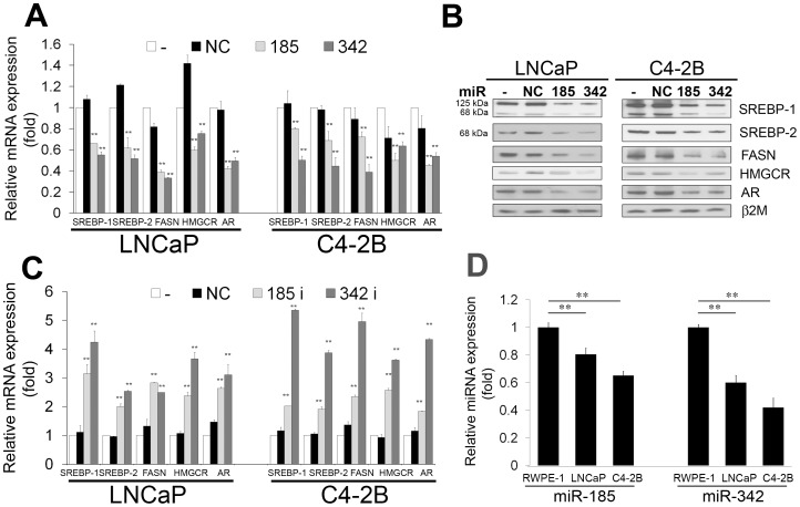
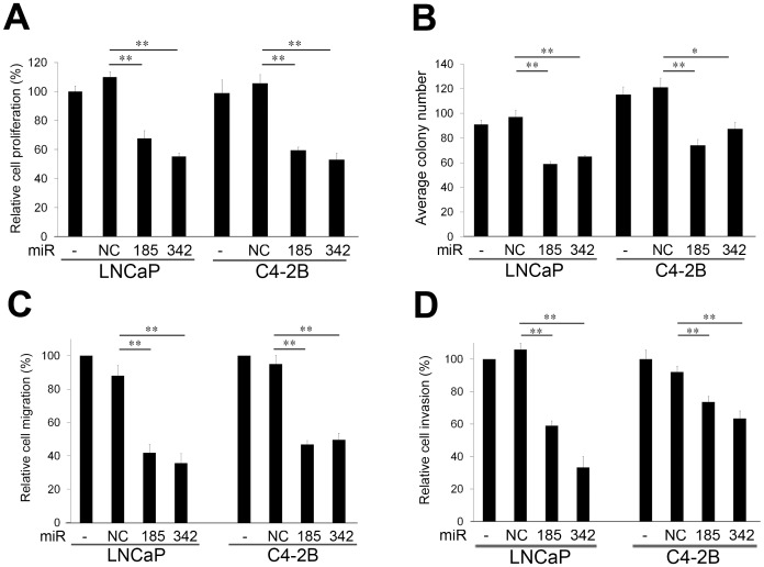
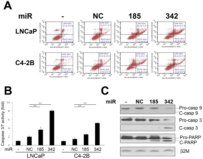
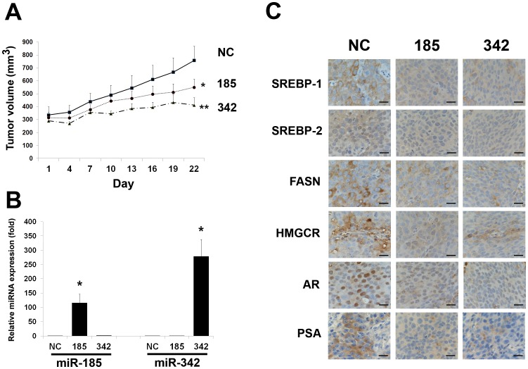
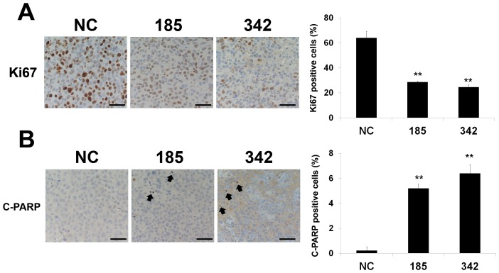
MicroRNA (miRNA or miR) inhibition of oncogenic related pathways has been shown to be a promising therapeutic approach for cancer. Aberrant lipid and cholesterol metabolism is involved in prostate cancer development and progression to end-stage disease. We recently demonstrated that a key transcription factor for lipogenesis, sterol regulatory element-binding protein-1 (SREBP-1), induced fatty acid and lipid accumulation and androgen receptor (AR) transcriptional activity, and also promoted prostate cancer cell growth and castration resistance. SREBP-1 was overexpressed in human prostate cancer and castration-resistant patient specimens. These experimental and clinical results indicate that SREBP-1 is a potential oncogenic transcription factor in prostate cancer. In this study, we identified two miRNAs, miR-185 and 342, that control lipogenesis and cholesterogenesis in prostate cancer cells by inhibiting SREBP-1 and 2 expression and down-regulating their targeted genes, including fatty acid synthase (FASN) and 3-hydroxy-3-methylglutaryl CoA reductase (HMGCR). Both miR-185 and 342 inhibited tumorigenicity, cell growth, migration and invasion in prostate cancer cell culture and xenograft models coincident with their blockade of lipogenesis and cholesterogenesis. Intrinsic miR-185 and 342 expression was significantly decreased in prostate cancer cells compared to non-cancerous epithelial cells. Restoration of miR-185 and 342 led to caspase-dependent apoptotic death in prostate cancer cells. The newly identified miRNAs, miR-185 and 342, represent a novel targeting mechanism for prostate cancer therapy.


Conflict of interest statement

Competing Interests: The authors have declared that no competing interests exist.

Figures

Figure 1
Figure 1. MiR-185 and 342 inhibition of SREBP mRNA and protein and expression patterns of miR-185 and 342 in prostate cancer cells.
A, Both miR-185 and 342 inhibited mRNA expression of SREBP-1, SREBP-2, FASN, HMGCR and AR in LNCaP and C4-2B prostate cancer cells determined by qRT-PCR. -: non-transfected; NC: miR-negative control. The relative mRNA expression (fold) was assigned as 1.0 in non-transfected cells. Data were normalized to 18S rRNA and represent the mean ± SD of three independent duplicate experiments. **, P < 0.005 significant differences from NC. B, MiR-185 and 342 inhibited precursor (125 kDa) and mature (68 kDa) forms of SREBP-1 and SREBP-2, FASN, HMGCR and AR expression in LNCaP and C4-2B cells assayed by Western blot. β2-microglobulin (β2M) was used as a loading control. C, MiR-185 and 342 inhibitors (antisense oligonucleotides against miR-185 and 342) increased SREBP-1, SREBP-2, FASN, HMGCR and AR expression in LNCaP and C4-2B cells determined by qRT-PCR. The relative mRNA expression (fold) was assigned as 1.0 in non-transfected cells. Data were normalized to 18S rRNA and represent the mean ± SD of three independent duplicate experiments. **, P < 0.005 significant differences from NC. D, Expression of intrinsic miR-185 and 342 in RWPE-1, LNCaP and C4-2B cells. qRT-PCR results showed that relative expression of miR-185 and 342 was significantly decreased in prostate cancer cells compared with normal/non-cancerous RWPE-1. Lower expression of both miRNAs was observed in aggressive C4-2B compared with LNCaP cells. The relative miRNA expression (fold) was assigned as 1.0 in RWPE-1 cells. **, P < 0.005 significant differences from RWPE-1. Data were normalized to RNU6B control and represent the mean ± SD of three independent experiments performed in quadruplicate.
Figure 2
Figure 2. MiR-185 and 342 suppress cell proliferation, clonogenicity, migration and invasion.
A, MiR-185 and 342 inhibited cell proliferation in LNCaP and C4-2B cells compared with non-transfected (−) and miR-negative control (NC) transfected cells 3 d following miRNA transfection. The relative cell proliferation (%) was assigned as 100% in non-transfected (−) cells. **, P < 0.005 significant differences from NC. Data represent the mean ± SD of two independent quadruplicate experiments. B, MiR-185 and 342 suppressed colony formation in LNCaP and C4-2B cells compared with the control groups after 14 d miRNA transfection. *, P < 0.05 and **, P < 0.005 significant differences from NC. Data represent the mean ± SD of two independent triplicate experiments. C, Cell migration and D, invasion were significantly decreased by miR-185 and 342 in LNCaP and C4-2B cells compared with the control groups. **, P < 0.005 significant differences from NC. Data represent the mean ± SD of two independent quadruplicate experiments.
Figure 3
Figure 3. MiR-185 and 342 induce caspase-dependent apoptotic death.
A, An Annexin V-FITC/PI staining apoptotic assay and flow cytometry were performed in LNCaP and C4-2B cells transfected with miRNAs. Both miR-185 and 342 increased the apoptotic cell fractions (both early and late apoptotic cell fractions; P<0.005) compared with the control groups. B, Caspase 3/7 activities were significantly increased by miR-185 and 342 in LNCaP and C4-2B cells. Caspase 3/7 activities (fold) were assigned as 1.0 in non-transfected (−) cells. **, P < 0.005 significant differences from NC. Data represent the mean ± SD of two independent triplicate experiments. C, MiR-185 and 342 decreased pro-caspase 9, 3 and PARP, and activated cleaved caspase 3 and PARP expression in LNCaP cells as assayed by Western blot. β2M was used as a loading control. C: cleaved.
Figure 4
Figure 4. Intratumoral delivery of miR-185 and 342 leads to regression of prostate tumors in a mouse xenograft model.
A, Subcutaneous C4-2B tumor growth was assayed by tumor volume after intratumoral delivery of miR-185, 342 or NC every 3 d for a 21-d treatment in mice. Both miR-185 and 342 significantly inhibited the growth of C4-2B tumors compared with NC-treated tumors. *, P<0.05; **, P<0.005 significant differences from NC-treated tumors (N = 5 for each group). B, The relative expression of miR-185 or 342 in C4-2B tumors. qRT-PCR results showed that the relative miR-185 or 342 levels were greatly increased in the subcutaneous C4-2B tumors injected with miR-185 or 342 compared with the control tumors. The relative miRNA expression (fold) was assigned as 1.0 in NC. *, P < 0.05 significant differences from NC. Data were normalized to RNU6B and represent the mean ± SD. C, IHC results showed that down-regulation of SREBP-1, SREBP-2, FASN, HMGCR, AR and PSA was observed in the miR-185 and 342-treated subcutaneous C4-2B tumors in comparison with the NC-treated tumors. Scale bars = 25 µm.
Figure 5
Figure 5. MiR-185 and 342 inhibit cell proliferation and induce apoptosis in subcutaneous xenografts.
Quantification of A, Ki67 (cell proliferation) and B, cleaved PARP (C-PARP; apoptosis) positive cells in subcutaneous C4-2B tumor specimens collected from the miR-NC, 185 and 342 treated groups. One hundred cells at 5 randomly selected areas were counted and positively staining cells were recorded. **, P < 0.005 significant differences from the control NC group. Data represent the mean ± SD. Arrowheads indicate the C-PARP positive cells. Scale bars = 100 µm.

References

    1. Bartel DP (2009) MicroRNAs: target recognition and regulatory functions. Cell 136: 215–233. - PMC - PubMed
    1. Bartel DP (2004) MicroRNAs: genomics, biogenesis, mechanism, and function. Cell 116: 281–297. - PubMed
    1. Fernandez-Hernando C, Suarez Y, Rayner KJ, Moore KJ (2011) MicroRNAs in lipid metabolism. Curr Opin Lipidol 22: 86–92. - PMC - PubMed
    1. Hime GR, Somers WG (2009) Micro-RNA mediated regulation of proliferation, self-renewal and differentiation of mammalian stem cells. Cell Adh Migr 3: 425–432. - PMC - PubMed
    1. Krutzfeldt J, Stoffel M (2006) MicroRNAs: a new class of regulatory genes affecting metabolism. Cell Metab 4: 9–12. - PubMed

Publication types

MeSH terms