. 2013 Aug 12;8(8):e71193.
doi: 10.1371/journal.pone.0071193. eCollection 2013.

The GPVI-Fc fusion protein Revacept reduces thrombus formation and improves vascular dysfunction in atherosclerosis without any impact on bleeding times

Affiliations

The GPVI-Fc fusion protein Revacept reduces thrombus formation and improves vascular dysfunction in atherosclerosis without any impact on bleeding times

Martin Ungerer et al. PLoS One. .

Abstract

Aims: Glycoprotein VI (GPVI) is a key platelet receptor which mediates plaque-induced platelet activation and consecutive atherothrombosis, but GPVI is also involved in platelet-mediated atheroprogression. Therefore, interference in GPVI-mediated platelet activation has the potential to combine short-term and long-term beneficial effects, specificity and safety especially regarding bleeding complications.

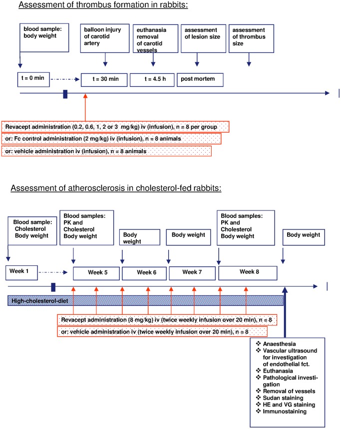
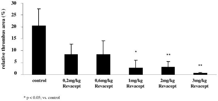
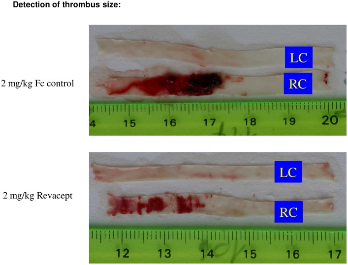
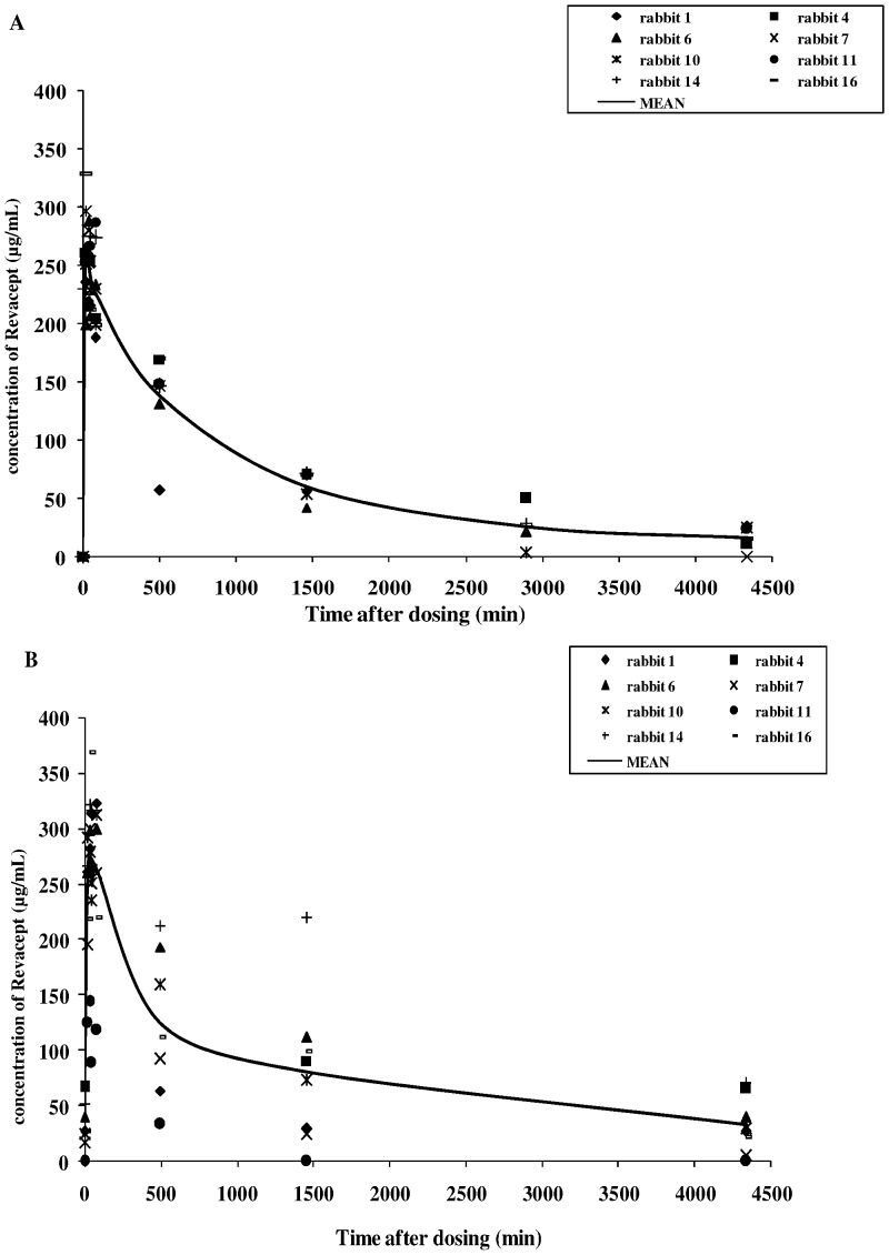
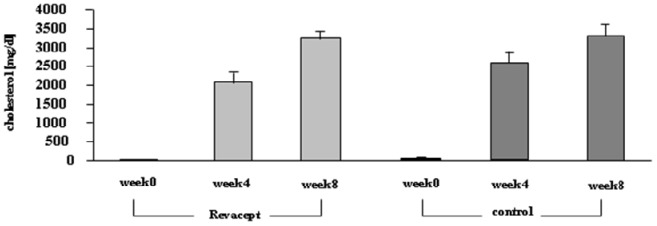
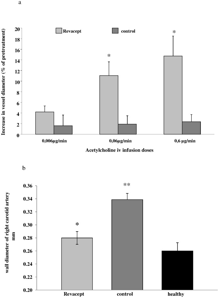
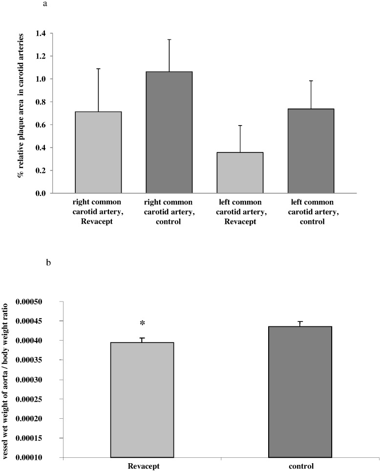
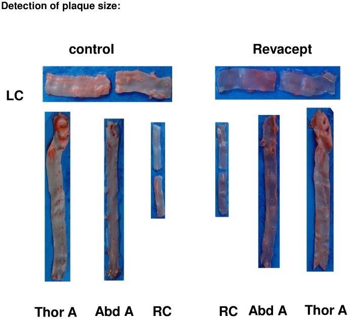
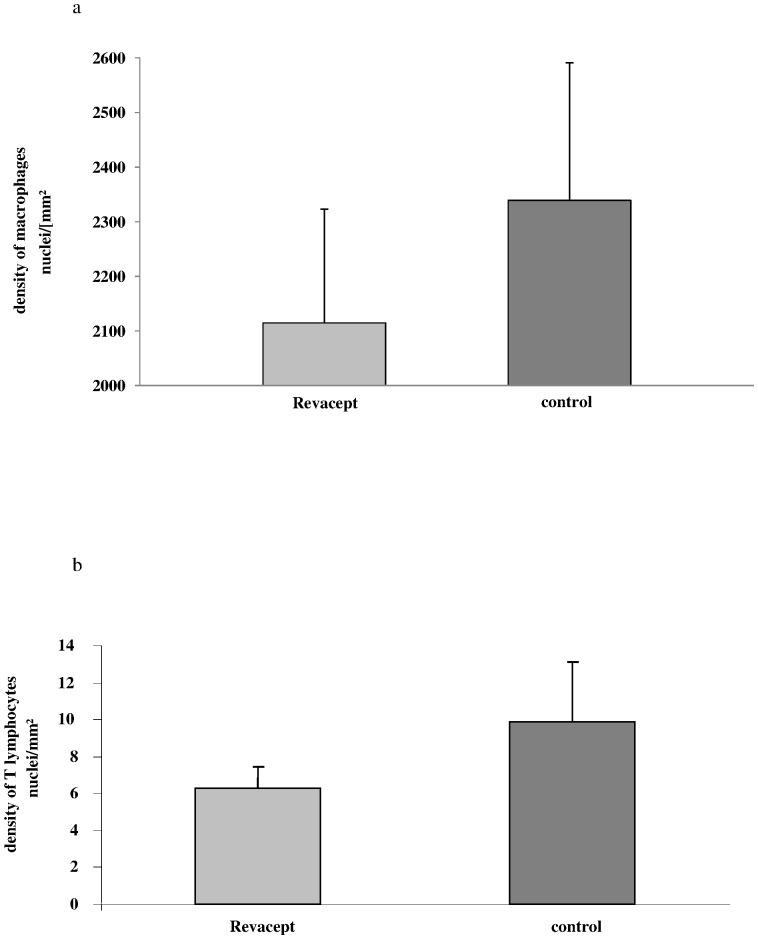
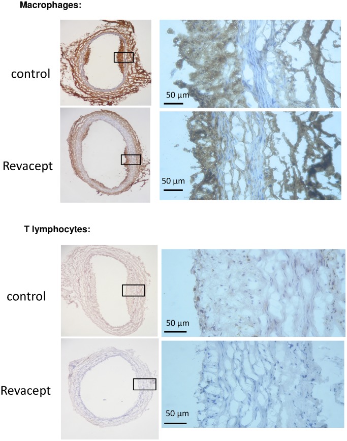
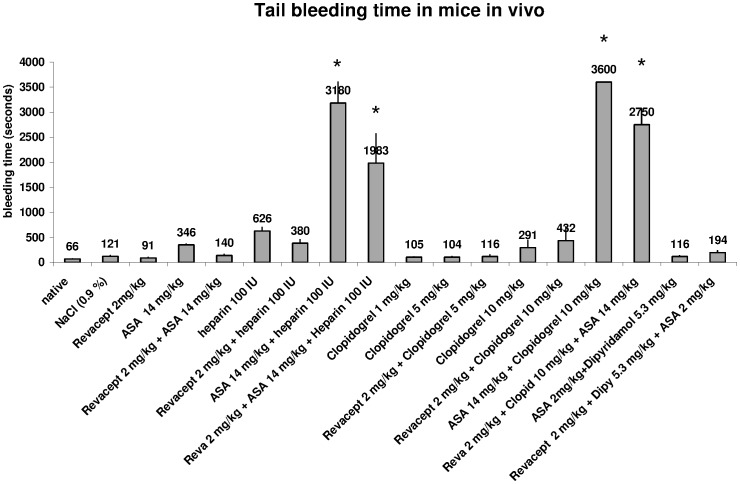
Methods and results: We investigated the effects of the soluble dimeric GPVI receptor fusion protein, Revacept, an antagonist of collagen-mediated platelet activation, in an animal model of atherosclerosis: twenty week old rabbits, which had been fed on a cholesterol-rich diet for 8 weeks, received Revacept (8 mg/kg) or control twice weekly for 4 weeks. Pharmacokinetics indicated a slight accumulation of the drug in the serum after repeated dosing of Revacept for 3 weeks. A significant improvement of endothelial dysfunction after 0.06 and 0.6 µg/min acetylcholine and a significant decrease of vessel wall thickening were found after Revacept treatment. Accordingly, aortic vessel weight was reduced, and plaque sizes, macrophage and T-cell invasion tended to be reduced in histological evaluations. Bleeding time was determined after tail clipping in mice. Revacept alone or in combination with widely used anti-platelet drugs revealed a high safety margin with no prolongation of bleeding times.

Conclusion: Repeated doses of Revacept led to a significant improvement of endothelial dysfunction and vascular morphology in atherosclerotic rabbits. Furthermore, no influence of Revacept on bleeding time alone or in combinations with various anti-platelet drugs was found in mice. Thus, the inhibition of collagen-mediated platelet interaction with the atherosclerotic endothelium by Revacept exerts beneficial effects on morphology and vascular function in vivo and seems to have a wide therapeutic window without influencing the bleeding time.


Conflict of interest statement

Competing Interests: Authors MU, ZL, SG, JV, HPH and GM are employed by, or hold shares of the biotech company advanceCOR GmbH - this company funded this study. MG is a co-founder of AdvanceCOR, owns shares of AdvanceCOR and is Professor at the Cardiology Department of the University of Tübingen. He further received honoraria payments from Lilly, Bristol-Myers Squibb and Bayer-Schering and is also consultant for Bayer-Schering. CB was an employee of the predecessor company, ProCorde GmbH, at the time of the study. Advancecor holds patents on and develops GPVI-Fc as a product for the treatment of patients with vascular diseases. There are no marketed products to declare. This does not alter the authors' adherence to all the PLOS ONE policies on sharing data and materials.

Figures

Figure 1
Figure 1. Study protocols of thrombus size detection after carotis artery lesion (upper panel), and of atherosclerosis in cholesterol-fed rabbits after eight weeks of drug administration (lower panel).
Figure 2
Figure 2. Effect of systemic delivery of increasing doses of Revacept in rabbits.
Thrombus formation was induced by balloon vascular injury. The thrombus size was evaluated histologically post mortem and is expressed as % of the total vascular lesion area. The mean ± SD of n = 8 experiments per group are shown. * indicates significant difference of p<0.05, and ** of p<0.01, versus controls (as determined by ANOVA).
Figure 3
Figure 3. Representative images of thrombi in carotid artery preparations (en face).
LC: left carotid artery; RC: right carotid artery.
Figure 4
Figure 4. Pharmacokinetic profile of Revacept in rabbits (A) on the first day of dosing (after 4 weeks of high cholesterol feeding), (B) after the last dosing of the twice weekly dosing period four weeks later.
The graphs show single animal and mean values of the Revacept group.
Figure 5
Figure 5. Serum cholesterol levels of the Revacept group and the control group before, 4 and 8 weeks after feeding a high-cholesterol diet (means ± SEM).
Figure 6
Figure 6. Endothelial dysfunction and arterial wall thickness was assessed in rabbits after 8 weeks of high cholesterol diet.
(a) Vascular ultrasound of the right common artery: The bars show increases of luminal diameter of the right common carotid artery [%] compared to baseline after infusion of three doses of acetylcholine in the Revacept and control groups. (b) Wall thickness of the right common carotid artery [mm] in rabbits. The mean ± SEM of 8 animals are shown. * indicates significant difference of p<0.05 versus cholesterol-fed, atherosclerotic control rabbits, and ** p<0.01 versus healthy rabbits (ANOVA).
Figure 7
Figure 7. Macroscopic and histological assessment of atherosclerosis in cholesterol-fed rabbits.
(a) Plaque size [%] was determined in macroscopic en face preparations after sudan red staining of the common carotid arteries. The relative lesion area was expressed as percentage of the total vessel area of the common carotid artery. (b) Vessel wet weight to body weight ratio was determined. The mean ± SEM of 8 animals are shown. * indicates significant difference of p<0.05 versus cholesterol-fed atherosclerotic control rabbits.
Figure 8
Figure 8. Representative images of plaque extensions in artery sections.
LC: left carotid artery; RC: right carotid artery, Thor A: thoracic aorta, Abd A: abdominal aorta.
Figure 9
Figure 9. The inflammation in the arterial wall was assessed by immune-histology in carotid artery sections of cholesterol fed rabbits.
(a) The density of macrophages was determined using specific anti-RAM antibodies.(b) The density of T-lymphatic cells was determined with specific anti CD 4 antibodies. The mean ± SEM of 8 animals are shown.
Figure 10
Figure 10. Representative immuno-histological images of macrophage and T lymphocyte stains of carotid artery sections.
Figure 11
Figure 11. Tail bleeding time was assessed after tail clipping in mice.
Revacept was investigated alone and in combination with existing drugs. The means ± SEM of n = 8 animals per group are demonstrated. Stars (*) indicate significant differences, p<0.05 versus native, untreated mice, or vs. mice which were only treated with 2 mg/kg Revacept (ANOVA).

References

    1. Massberg S, Konrad I, Bültmann A, Schulz C, Münch G, et al. (2004) Soluble glycoprotein VI dimer inhibits platelet adhesion and aggregation to the injured vessel wall in vivo. FASEB J 18(2): 397–399. - PubMed
    1. Massberg S, Gawaz M, Grüner S, Schulte V, Konrad I, et al. (2003) A crucial role of glycoprotein VI for platelet recruitment to the injured arterial wall in vivo. J Exp Med 197(1): 41–49. - PMC - PubMed
    1. Reininger AJ, Bernlochner I, Penz SM, Ravanat C, Smethurst P, et al. (2010) A 2-step mechanism of arterial thrombus formation induced by human atherosclerotic plaques. J Am Coll Cardiol 55(11): 1147–1158. - PubMed
    1. Massberg S, Brand K, Grüner S, Page S, Müller E, et al. (2002) A critical role of platelet adhesion in the initiation of atherosclerotic lesion formation. J Exp Med 196(7): 887–896. - PMC - PubMed
    1. Kastrati A, Neumann FJ, Mehilli J, Byrne RA, Iijima R, et al. (2008) Bivalirudin versus unfractionated heparin during percutaneous coronary intervention. N Engl J Med 359(7): 688–696. - PubMed

MeSH terms