. 2013 Aug 7;8(8):e71316.
doi: 10.1371/journal.pone.0071316. eCollection 2013.

Rhinovirus 3C protease facilitates specific nucleoporin cleavage and mislocalisation of nuclear proteins in infected host cells

Affiliations

Rhinovirus 3C protease facilitates specific nucleoporin cleavage and mislocalisation of nuclear proteins in infected host cells

Erin J Walker et al. PLoS One. .

Abstract

Human Rhinovirus (HRV) infection results in shut down of essential cellular processes, in part through disruption of nucleocytoplasmic transport by cleavage of the nucleoporin proteins (Nups) that make up the host cell nuclear pore. Although the HRV genome encodes two proteases (2A and 3C) able to cleave host proteins such as Nup62, little is known regarding the specific contribution of each. Here we use transfected as well as HRV-infected cells to establish for the first time that 3C protease is most likely the mediator of cleavage of Nup153 during HRV infection, while Nup62 and Nup98 are likely to be targets of HRV2A protease. HRV16 3C protease was also able to elicit changes in the appearance and distribution of the nuclear speckle protein SC35 in transfected cells, implicating it as a key mediator of the mislocalisation of SC35 in HRV16-infected cells. In addition, 3C protease activity led to the redistribution of the nucleolin protein out of the nucleolus, but did not affect nuclear localisation of hnRNP proteins, implying that complete disruption of nucleocytoplasmic transport leading to relocalisation of hnRNP proteins from the nucleus to the cytoplasm in HRV-infected cells almost certainly requires 2A in addition to 3C protease. Thus, a specific role for HRV 3C protease in cleavage and mislocalisation of host cell nuclear proteins, in concert with 2A, is implicated for the first time in HRV pathogenesis.


Conflict of interest statement

Competing Interests: The authors have declared that no competing interests exist.

Figures

Figure 1
Figure 1. Specific nucleoporins are degraded in HRV16-infected cells.
(A) Ohio-HeLa cells were infected without (mock) or with HRV16 (MOI of 1) and cells lysed using RIPA buffer containing protease and phosphatase inhibitors at the time points shown. Cell lysates were subjected to SDS-PAGE on 4–20% gradient gels and Western analysis using the indicated primary antibodies/horseradish peroxidise-conjugated secondary antibodies and enhanced chemiluminescence (Perkin Elmer). The specificity of the antibodies is indicated on the left. Bands corresponding to 3C, 3CD’ and 3CD are indicated on the right. p.i. - post-infection. (B) Results for densitometric analysis of FG-Nup protein bands (left) and non-FG-Nups (right) such as those shown in (A), where data were normalised to the corresponding values for tubulin, relative to the corresponding values for the mock sample. Densitometric analyses were performed using Image J; values represent the mean (± SD) from two independent experiments.
Figure 2
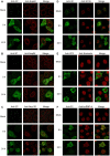
Figure 2. HRV16 infection leads to mislocalisation of nuclear proteins.
Ohio-HeLa cells grown on coverslips were infected without (mock) or with HRV16 as per Figure 1; cells were fixed at the indicated times and permeabilized, and then probed with the indicated pairs of primary antibodies, followed by Alexa 488 and Alexa-568 conjugated secondary antibodies. Fluorescence was imaged by CLSM (see Materials and Methods). In each panel, images on the left depict localisation of HRV16 proteins (green channel) and the images in the middle depict localisation of cellular proteins (red channel), with the merged image on the right.
Figure 3
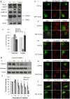
Figure 3. Active 3C protease is sufficient to degrade nuclear proteins/nucleoporins in transfected cells.
(Ai) COS-7 cells transfected to express either GFP-3C or GFP-3Cinac were trypsinized 18 h after transfection, harvested in ice-cold PBS and FACS sorted to collect GFP-expressing cells. Cells were then lysed using RIPA buffer plus protease and phosphatase inhibitors and lysates were subjected to Western analysis as per Figure 1A; untransfected cells were lysed similarly and used as control. The antibodies used are shown on the left of the figure. The arrow in the nucleolin blot denotes a clear cleavage product, with the approximate molecular weight (kDa) indicated on the right. (Aii) Results for densitometric analysis of nucleolin and Nup153 protein bands such as those shown in Figure 3Ai, where data were normalised to the corresponding value of tubulin, relative to the corresponding value for the control sample. Densitometric analyses were performed using ImageJ; values represent the mean (+ SD) from two independent experiments (Bi) Ohio-Hela whole cell lysates were incubated with 4 units of HRV14 3C protease at 37°C for the indicated incubation times, subsequent to SDS-PAGE on 10% gels and Western analysis as per Figure 1A. (Bii) Plot of densitometric analysis of protein bands in (Bi), where data were normalised to the corresponding values for tubulin, relative to 0 h samples. Densitometric analyses were performed using Image J and values were the mean of two different experiments (± SD). (C) COS-7 cells transfected to express either GFP-3C or GFP-3Cinac were fixed and permeabilized 18 h post-transfection, and immunostained as described in Figure 2 with the indicated primary and Alexa-568 conjugated secondary antibodies. Fluorescence was imaged by CLSM (see Materials and Methods). In each panel, images on the left depict localisation of HRV16 proteins (green channel) and the images in the middle depict localisation of cellular proteins (red channel), with the merged image on the right.
Figure 4
Figure 4. Schematic representation of proposed FG-Nup cleavage by 2A and 3C proteases during HRV16 infection.
During infection with HRV16, the initial cleavage event within the nuclear pore is mediated by 2A protease and leads to Nup98 cleavage (1). Cleavage of Nup153 by 3C protease occurs at approximately 6–9 h p.i. correlating with the localisation of 3C in the nucleus (2). Mislocalisation of SC35 from nuclear speckles to the nucleus and cytoplasm and mislocalisation of hnRNP-A1 from the nucleus to the cytoplasm is detected at 6 h p.i. (3). Cleavage of Nup62 by 2A protease occurs later, between 9–24 h p.i. (4). Nups that were cleaved during infection are shown in red type while Nups that were unaffected during infection are shown in black type.

References

    1. Johnston SL, Pattemore PK, Sanderson G, Smith S, Lampe F, et al. (1995) Community study of role of viral infections in exacerbations of asthma in 9–11 year old children. BMJ 310: 1225–1229. - PMC - PubMed
    1. Kaur B, Anderson HR, Austin J, Burr M, Harkins LS, et al. (1998) Prevalence of asthma symptoms, diagnosis, and treatment in 12–14 year old children across Great Britain (international study of asthma and allergies in childhood, ISAAC UK). BMJ 316: 118–124. - PMC - PubMed
    1. Racaniello VR (2001) Picornaviridae: the viruses and their replication. In: Knipe DM, Howley PM, editors. Fields Virology, 4th Edition. Philadelphia, PA: Lippincott/Raven. 685–722.
    1. Brunel F, Zakin MM, Buc H, Buckle M (1996) The Polypyrimidine Tract Binding (PTB) Protein Interacts With Single-Stranded DNA in a Sequence-Specific Manner. Nucleic Acids Research 24: 1608–1615. - PMC - PubMed
    1. Nagy PD, Wang RY, Pogany J, Hafren A, Makinen K (2011) Emerging picture of host chaperone and cyclophilin roles in RNA virus replication. Virology 411: 374–382. - PubMed

Publication types

MeSH terms

LinkOut - more resources