

Impaired blood dendritic cell numbers and functions after aneurysmal subarachnoid hemorrhage
- PMID: 23951210
- PMCID: PMC3739744
- DOI: 10.1371/journal.pone.0071639
Impaired blood dendritic cell numbers and functions after aneurysmal subarachnoid hemorrhage
Abstract
Previous presentation: Portions of this study were presented at the Annual Congress of Société Française d'Anesthésie et de Réanimation in Paris, September 2012.
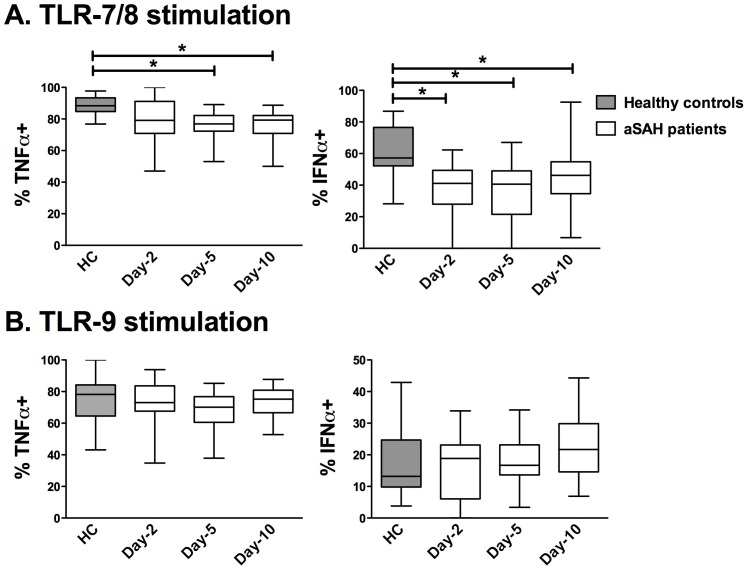
Background: Toll-like receptor (TLR) agonists are promising therapy for the prevention of nosocomial infections in critical ill patients. We aimed to analyze the TLR-reactivity of circulating dendritic cells (DC) as assessed by cytokine production after an ex vivo challenge with TLR agonists in aneurysmal subarachnoid hemorrhage (SAH) patients.
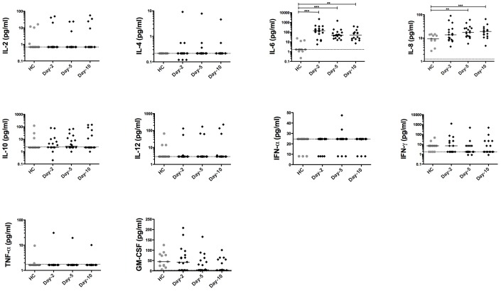
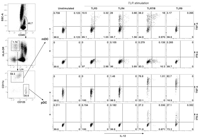
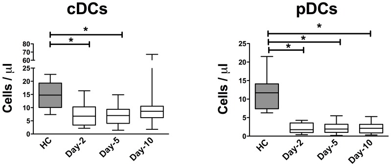
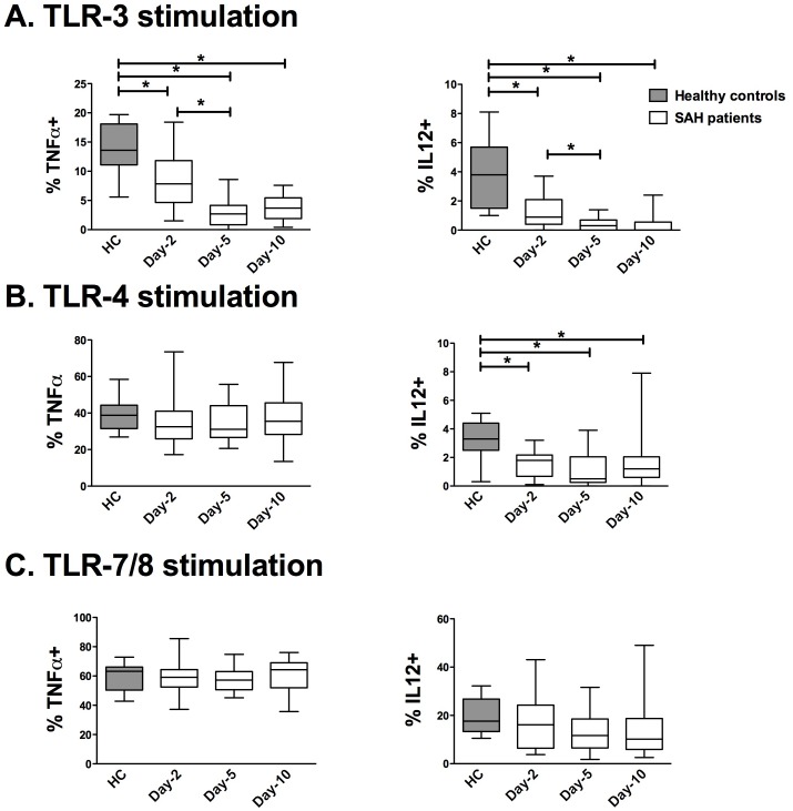
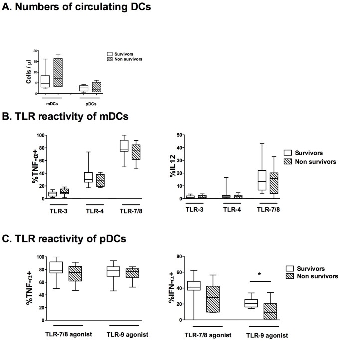
Methods and findings: A single-center prospective observational study took place in one intensive care unit of a teaching hospital. Blood samples were harvested on days 2, 5 and 10 in 21 severe SAH patients requiring mechanical ventilation and 17 healthy controls. DC production of cytokines (Tumour Necrosis Factor, TNF-α; Interleukin, IL-12; and Interferon, IFN-α) was assessed by intracellular immunostaining on TLR-3, 4, 7/8 and 9 stimulations. SAH patients had decreased numbers of blood myeloid (mDCs) and plasmacytoid DCs (pDCs) on days 2, 5 and 10. Compared with the healthy controls, the frequency of mDCs producing TNF-α after TLR-3 stimulation was decreased in the SAH patients. The frequency of myeloid DCs producing IL-12 after TLR-3 and 4 stimulations was also decreased in the SAH patients. In contrast, the mDCs response to TLR-7/8 was not impaired in the SAH patients. The frequency of pDCs producing TNF-α(+) and IFN-α(+) on TLR-7/8 stimulation were reduced at all of the tested times in the SAH patients, whereas reactivity to TLR-9 was preserved. On day 2, the pDCs from non-survivor patients (n=8) had a decreased ability to produce IFN-α on TLR-9 stimulation compared with the survivors.
Conclusions: These data suggest functional abnormalities of circulating pDCs and mDCs that could be important for immunomodulation after SAH.
Conflict of interest statement
Figures
Similar articles
-
Toll-like receptor ligands modulate dendritic cells to augment cytomegalovirus- and HIV-1-specific T cell responses.J Immunol. 2003 Oct 15;171(8):4320-8. doi: 10.4049/jimmunol.171.8.4320. J Immunol. 2003. PMID: 14530357
-
Age-associated decrease in TLR function in primary human dendritic cells predicts influenza vaccine response.J Immunol. 2010 Mar 1;184(5):2518-27. doi: 10.4049/jimmunol.0901022. Epub 2010 Jan 25. J Immunol. 2010. PMID: 20100933 Free PMC article.
-
Hydroxychloroquine is associated with impaired interferon-alpha and tumor necrosis factor-alpha production by plasmacytoid dendritic cells in systemic lupus erythematosus.Arthritis Res Ther. 2012 Jun 27;14(3):R155. doi: 10.1186/ar3895. Arthritis Res Ther. 2012. PMID: 22734582 Free PMC article.
-
Plasmacytoid DCs, exposed to TSLP in synergy with TLR ligands, acquire significant potential towards Th2 polarization.Med Sci Monit Basic Res. 2013 Dec 13;19:291-9. doi: 10.12659/msmbr.889791. Med Sci Monit Basic Res. 2013. PMID: 24335833 Free PMC article.
-
Fatigue, Toll-Like Receptor 4, and Pro-Inflammatory Cytokines in Adults With Subarachnoid Hemorrhage: A 6-Month Longitudinal Study.Biol Res Nurs. 2024 Apr;26(2):192-201. doi: 10.1177/10998004231203257. Epub 2023 Oct 3. Biol Res Nurs. 2024. PMID: 37788710 Review.
Cited by
-
Toll-Like Receptor Signaling Pathways: Novel Therapeutic Targets for Cerebrovascular Disorders.Int J Mol Sci. 2021 Jun 7;22(11):6153. doi: 10.3390/ijms22116153. Int J Mol Sci. 2021. PMID: 34200356 Free PMC article. Review.
-
Immune modulation after traumatic brain injury.Front Med (Lausanne). 2022 Dec 1;9:995044. doi: 10.3389/fmed.2022.995044. eCollection 2022. Front Med (Lausanne). 2022. PMID: 36530909 Free PMC article. Review.
-
Trauma-Induced Damage-Associated Molecular Patterns-Mediated Remote Organ Injury and Immunosuppression in the Acutely Ill Patient.Front Immunol. 2018 Jun 15;9:1330. doi: 10.3389/fimmu.2018.01330. eCollection 2018. Front Immunol. 2018. PMID: 29963048 Free PMC article. Review.
-
Contribution of Dendritic Cell Responses to Sepsis-Induced Immunosuppression and to Susceptibility to Secondary Pneumonia.Front Immunol. 2018 Nov 13;9:2590. doi: 10.3389/fimmu.2018.02590. eCollection 2018. Front Immunol. 2018. PMID: 30483258 Free PMC article. Review.
-
The role of Toll-like receptor signaling pathways in cerebrovascular disorders: the impact of spreading depolarization.J Neuroinflammation. 2020 Apr 7;17(1):108. doi: 10.1186/s12974-020-01785-6. J Neuroinflammation. 2020. PMID: 32264928 Free PMC article. Review.
References
-
- Bederson JB, Connolly ES, Batjer HH, Dacey RG, Dion JE, et al. (2009) Guidelines for the management of aneurysmal subarachnoid hemorrhage: a statement for healthcare professionals from a special writing group of the Stroke Council, American Heart Association. Stroke 40: 994–1025 doi:10.1161/STROKEAHA.108.191395 - DOI - PubMed
-
- Dorhout Mees SM, Algra A, Vandertop WP, van Kooten F, Kuijsten HAJM, et al. (2012) Magnesium for aneurysmal subarachnoid haemorrhage (MASH-2): a randomised placebo-controlled trial. Lancet 380: 44–49 doi:10.1016/S0140-6736(12)60724-7 - DOI - PMC - PubMed
-
- Stegmayr B, Eriksson M, Asplund K (2004) Declining Mortality From Subarachnoid Hemorrhage: Changes in Incidence and Case Fatality From 1985 Through 2000. Stroke 35: 2059–2063 doi:10.1161/01.STR.0000138451.07853.b6 - DOI - PubMed
-
- Sarrafzadeh A, Schlenk F, Meisel A, Dreier J, Vajkoczy P, et al. (2011) Immunodepression after aneurysmal subarachnoid hemorrhage. Stroke 42: 53–58 doi:10.1161/STROKEAHA.110.594705 - DOI - PubMed
-
- Meisel C, Schwab JM, Prass K, Meisel A, Dirnagl U (2005) Central nervous system injury-induced immune deficiency syndrome. Nat Rev Neurosci 6: 775–786 doi:10.1038/nrn1765 - DOI - PubMed
Publication types
MeSH terms
Substances
LinkOut - more resources
Full Text Sources
Other Literature Sources
