

The G-protein coupled estrogen receptor (GPER/GPR30) is a gonadotropin receptor dependent positive prognosticator in ovarian carcinoma patients
- PMID: 23951246
- PMCID: PMC3739730
- DOI: 10.1371/journal.pone.0071791
The G-protein coupled estrogen receptor (GPER/GPR30) is a gonadotropin receptor dependent positive prognosticator in ovarian carcinoma patients
Abstract
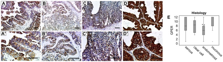
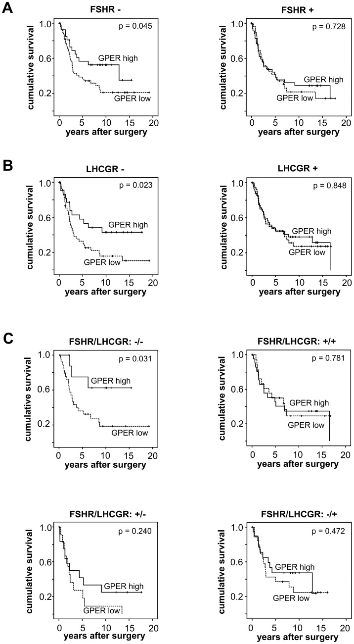
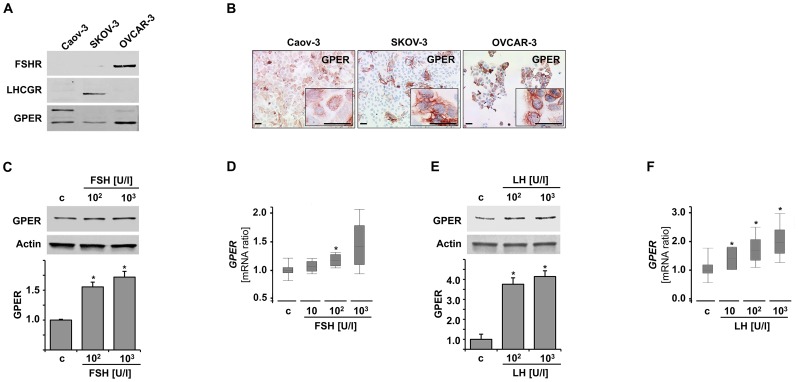
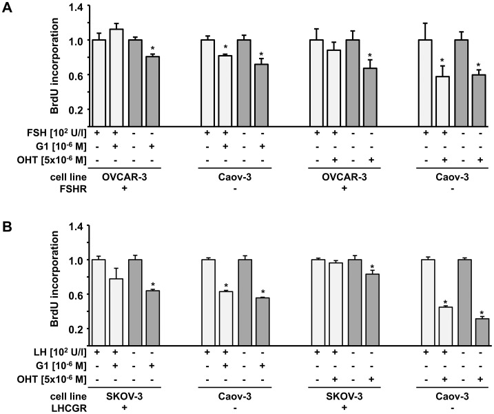
Follicle stimulating hormone receptor (FSHR) and luteinizing hormone receptor (LHCGR) were demonstrated to impact upon survival of patients suffering from epithelial ovarian cancer (EOC). Though structure wise the G-protein coupled estrogen receptor (GPER/GPR30) is related to FSHR/LHCGR, its prognostic impact in EOC remains controversial. We recently found that FSHR negative patients represent a specific EOC subgroup that may behave differently in respect to both treatment response and prognosis. Hence, the current study aimed to analyze how GPER may interact with the FSHR/LHCGR system in EOC and whether the prognostic significance of GPER in EOC cases (n=151) may be dependent on the FSHR/LHCGR immunophenotype of the tumor. Ovarian cancer cell lines were used to study how FSH and LH regulate GPER and whether GPER activation differentially affects in vitro cell proliferation in presence/absence of activated FSHR/LHCGR. In EOC tissue, GPER correlated with FSHR/LHCGR and was related to prolonged overall survival only in FSHR/LHCGR negative patients. Although GPER was found to be specifically induced by LH/FSH, GPER agonists (4-Hydroxy-Tamoxifen, G1) reduced EOC cell proliferation only in case of LH/FSH unstimulated pathways. To the same direction, only patients characterized as LHCGR/FSHR negative seem to gain from GPER in terms of survival. Our combined tissue and in vitro results support thus the hypothesis that GPER activation could be of therapeutic benefit in LHCGR/FSHR negative EOC patients. Further studies are needed to evaluate the impact of GPER activation on a clinical scheme.
Conflict of interest statement
Figures

Similar articles
-
Reduced Gonadotrophin Receptor Expression Is Associated with a More Aggressive Ovarian Cancer Phenotype.Int J Mol Sci. 2020 Dec 23;22(1):71. doi: 10.3390/ijms22010071. Int J Mol Sci. 2020. PMID: 33374698 Free PMC article.
-
G Protein-Coupled Estrogen Receptor Correlates With Dkk2 Expression and Has Prognostic Impact in Ovarian Cancer Patients.Front Endocrinol (Lausanne). 2021 Feb 19;12:564002. doi: 10.3389/fendo.2021.564002. eCollection 2021. Front Endocrinol (Lausanne). 2021. PMID: 33679613 Free PMC article.
-
Disruption of Zebrafish Follicle-Stimulating Hormone Receptor (fshr) But Not Luteinizing Hormone Receptor (lhcgr) Gene by TALEN Leads to Failed Follicle Activation in Females Followed by Sexual Reversal to Males.Endocrinology. 2015 Oct;156(10):3747-62. doi: 10.1210/en.2015-1039. Epub 2015 May 20. Endocrinology. 2015. PMID: 25993524
-
Follicle stimulating hormone receptor (FSHR) antagonist and epithelial ovarian cancer (EOC).J Exp Ther Oncol. 2007;6(3):201-4. J Exp Ther Oncol. 2007. PMID: 17552360 Review.
-
Epigenetic modifications of gonadotropin receptors can regulate follicular development.Anim Reprod Sci. 2024 Sep;268:107534. doi: 10.1016/j.anireprosci.2024.107534. Epub 2024 Jun 13. Anim Reprod Sci. 2024. PMID: 39047429 Review.
Cited by
-
Nuclear receptor co-repressor NCOR2 and its relation to GPER with prognostic impact in ovarian cancer.J Cancer Res Clin Oncol. 2023 Sep;149(11):8719-8728. doi: 10.1007/s00432-023-04708-z. Epub 2023 May 2. J Cancer Res Clin Oncol. 2023. PMID: 37131060 Free PMC article.
-
Downregulation of G Protein-Coupled Estrogen Receptor (GPER) is Associated with Reduced Prognosis in Patients with Gastric Cancer.Med Sci Monit. 2019 Apr 27;25:3115-3126. doi: 10.12659/MSM.913634. Med Sci Monit. 2019. PMID: 31028714 Free PMC article.
-
Estrogen signaling crosstalk: Implications for endocrine resistance in ovarian cancer.J Steroid Biochem Mol Biol. 2014 Sep;143:160-73. doi: 10.1016/j.jsbmb.2014.02.010. Epub 2014 Feb 22. J Steroid Biochem Mol Biol. 2014. PMID: 24565562 Free PMC article. Review.
-
The Role of Endocrine G Protein-Coupled Receptors in Ovarian Cancer Progression.Front Endocrinol (Lausanne). 2017 Apr 7;8:66. doi: 10.3389/fendo.2017.00066. eCollection 2017. Front Endocrinol (Lausanne). 2017. PMID: 28439256 Free PMC article. Review.
-
Follicle-Stimulating Hormone Receptor Expression and Its Potential Application for Theranostics in Subtypes of Ovarian Tumors: A Systematic Review.Cancers (Basel). 2024 Mar 13;16(6):1140. doi: 10.3390/cancers16061140. Cancers (Basel). 2024. PMID: 38539473 Free PMC article. Review.
References
-
- Karagol H, Saip P, Uygun K, Caloglu M, Eralp Y, et al. (2007) The efficacy of tamoxifen in patients with advanced epithelial ovarian cancer. Med Oncol 24: 39–43. - PubMed
-
- Wagner U, du Bois A, Pfisterer J, Huober J, Loibl S, et al. (2007) Gefitinib in combination with tamoxifen in patients with ovarian cancer refractory or resistant to platinum-taxane based therapy–a phase II trial of the AGO Ovarian Cancer Study Group (AGO-OVAR 2.6). Gynecol Oncol 105: 132–137. - PubMed
-
- Crespo P, Xu N, Simonds WF, Gutkind JS (1994) Ras-dependent activation of MAP kinase pathway mediated by G-protein beta gamma subunits. Nature 369: 418–420. - PubMed
Publication types
MeSH terms
Substances
LinkOut - more resources
Full Text Sources
Other Literature Sources
Medical
