. 2013 Aug 12;8(8):e71814.
doi: 10.1371/journal.pone.0071814. eCollection 2013.

Mesenchymal stem cell conditioning promotes rat oligodendroglial cell maturation

Affiliations

Mesenchymal stem cell conditioning promotes rat oligodendroglial cell maturation

Janusz Joachim Jadasz et al. PLoS One. .

Abstract

Oligodendroglial progenitor/precursor cells (OPCs) represent the main cellular source for the generation of new myelinating oligodendrocytes in the adult central nervous system (CNS). In demyelinating diseases such as multiple sclerosis (MS) myelin repair activities based on recruitment, activation and differentiation of resident OPCs can be observed. However, the overall degree of successful remyelination is limited and the existence of an MS-derived anti-oligodendrogenic milieu prevents OPCs from contributing to myelin repair. It is therefore of considerable interest to understand oligodendroglial homeostasis and maturation processes in order to enable the development of remyelination therapies. Mesenchymal stem cells (MSC) have been shown to exert positive immunomodulatory effects, reduce demyelination, increase neuroprotection and to promote adult neural stem cell differentiation towards the oligodendroglial lineage. We here addressed whether MSC secreted factors can boost the OPC's oligodendrogenic capacity in a myelin non-permissive environment. To this end, we analyzed cellular morphologies, expression and regulation of key factors involved in oligodendroglial fate and maturation of primary rat cells upon incubation with MSC-conditioned medium. This demonstrated that MSC-derived soluble factors promote and accelerate oligodendroglial differentiation, even under astrocytic endorsing conditions. Accelerated maturation resulted in elevated levels of myelin expression, reduced glial fibrillary acidic protein expression and was accompanied by downregulation of prominent inhibitory differentiation factors such as Id2 and Id4. We thus conclude that apart from their suggested application as potential anti-inflammatory and immunomodulatory MS treatment, these cells might also be exploited to support endogenous myelin repair activities.


Conflict of interest statement

Competing Interests: The authors have declared that no competing interests exist.

Figures

Figure 1
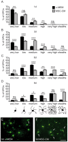
Figure 1. MSC-CM stimulates morphological maturation of OPCs.
Mesenchymal stem cell conditioned medium (MSC-CM) accelerates maturation of cultured oligodendroglial progenitors cells. We identified six distinct morphologies (see bottom), from a very low number of processes in progenitor cells to multiple process-bearing cells (low, medium, high) to mature cells with a very high degree of arborisation or flattened appearance (sheaths). (A–D) Analysis of OPC morphology distribution revealed an MSC-CM-dependent shift towards more mature cells (black bars α-MEM treated cells; grey bars MSC-CM stimulated cells) after one (A), three (B), six (C) and nine days (D). (E,F) Representative citrine labelled OPCs revealing advanced morphologies and promoted sheath formation (arrows) upon MSC-CM stimulation after nine days. Data are shown as mean values ± SEM derived from n = 5 experiments. t-test (ns: not significant, * P<0.05; ** P<0.01; *** P<0.001). Scale bar: 50 µm.
Figure 2
Figure 2. MSC-CM leads to enhanced early myelin expression.
Determination of transcript levels by means of quantitative real-time RT-PCR. (A) Upregulation of ceramide galactosyltransferase (CGT) expression was detected after three, six and nine days in culture, (B) whereas gene expression levels of CNPase were elevated at every measured time point. Glyceraldehyde-3-phosphate dehydrogenase (GAPDH) expression was used as reference. (C) In addition, an increased percentage of CNPase-expressing OPCs was observed among MSC-CM treated cells as compared to cells grown in α-MEM. Significant differences were detected from day three onwards. (D–E’’’) Representative immunofluorescent stainings of CNPase expressing OPCs at all four-time points of investigation. Data are shown as mean values ± SEM and derive from n = 8 (CGT), n = 8 (CNPase, q-RT-PCR) and n = 4 (CNPase, immunostainings) experiments. t-test (* P<0.05; ** P<0.01; *** P<0.001). Scale bars: 50 µm.
Figure 3
Figure 3. MSC-CM enhances myelin basic protein expression.
(A) Gene expression levels of MBP are up-regulated after one, three, six and nine days in MSC-CM. (B) Determination of the degree of MBP-positive OPCs as revealed by immunostainings show a significantly increased number after MSC-CM treatment for six and nine days. (C) Western blot analysis shows increased MBP protein levels after six days in mesenchymal stem cell conditioned medium. GAPDH was used as internal reference. (D–E’’’) Representative immunofluorescent stainings of MBP expressing OPCs at all four time points of investigation. Data are shown as mean values ± SEM derived from n = 6 (q-RT-PCR) and n = 3 (immunostainings, Western Blot) experiments. t-test (* P<0.05; ** P<0.01, *** P<0.001). Scale bars: 50 µm.
Figure 4
Figure 4. MSC-CM treatment enhances OPC proliferation, while cell death rate remains low.
(A) Anti-Ki67 immunofluorescent stainings and evaluation of positive cells revealed a higher proliferation rate under MSC-CM treatment as compared to α-MEM, whereas (B) cell death rates remained low under both conditions with a minor significant survival effect upon MSC-CM treatment at six days. Data are shown as mean values ± SEM derived from n = 4 experiments. t-test (ns: not significant, * P<0.05; **P<0.01).
Figure 5
Figure 5. MSC-CM dependent regulation of early OPC markers.
(A) Determination of the degree of A2B5 positive cells as revealed by immunofluorescence stainings. α-MEM and MSC-CM were given to the cells after four hours. (B) Determination of the degree of weak and strong Olig2 expressing cells during the course of α-MEM and MSC-CM stimulation up to nine days. (C–H’) Representative immunofluorescent stainings of Olig2 expressing OPCs at all three time points. Arrowheads mark weak expressing cells, whereas arrows point to strong expressors. Data are shown as mean values ± SEM derived from n = 3 (A2B5) and n = 4 (Olig2) experiments. t-test (ns: not significant, * P<0.05; **P<0.01; *** P<0.001). Scale bar: 50 µm.
Figure 6
Figure 6. Downregulation of GFAP expression following stimulation with MSC-CM.
OPCs were plated for four hours in SATO medium before changing the medium to control (α-MEM) or to mesenchymal stem cell conditioned medium (MSC-CM) and analysis after one, three, six and nine days. (A) Consistently decreased GFAP transcript levels were detected at every time point using quantitative real-time RT-PCR. (B) Determination of the degree of GFAP-positive OPCs revealed increasing numbers among α-MEM treated cells whereas MSC-CM stimulation stabilized low GFAP expression levels. (C–D’’’) Representative anti-GFAP immunofluorescent stainings. Data are shown as mean values ± SEM derived from n = 3 for both, q-RT-PCR as well as immunostaining experiments. t-test (*** P<0.001). Scale bars: 50 µm.
Figure 7
Figure 7. Serum reduced conditioned medium.
Determination of transcript levels by means of quantitative real-time RT-PCR. Investigation of GFAP, Id2 and Id4 gene expression levels in α-MEM versus MSC-CM containing 10% FBS (A,C,E). Investigation of GFAP, Id2 and Id4 gene expression levels in α-MEM versus MSC-CM containing 1% FBS (B,D,F). Glyceraldehyde-3-phosphate dehydrogenase (GAPDH) expression was used as reference gene; data are shown as mean values ± SEM derived from n = 7 for Id2, Id4 and n = 3 for GFAP (A,C,E) and n = 3 (B,D,F) experiments. t-test (ns: not significant, * P<0.05; **P<0.01; *** P<0.001).
Figure 8
Figure 8. HGF does not mimic MSC-CM mediated cellular effects.
(A) Determination of HGF protein content in stimulation media by means of ELISA revealing a robust increase upon MSC conditioning. (B) No difference regarding CNPase positivity was observed among OPCs grown in α-MEM in the absence or presence of recombinant HGF, whereas MSC-CM reproducibly increased CNPase expression. (C–E) Determination of transcript levels by means of quantitative real-time RT-PCR. No significant differences regarding Id2, Id4 and GFAP transcript levels were observed among OPCs grown in α-MEM in the absence or presence of recombinant HGF. MSC-CM treatment significantly reduced transcript levels of all three genes at all time points. (F–K) Anti-HGF antibody mediated depletion experiments revealed no effect on astrocyte (GFAP, Id2, Id4) and oligodendroglial/myelin (CGT, MBP, CNPase) gene expression levels. Glyceraldehyde-3-phosphate dehydrogenase (GAPDH) expression was used as reference gene. All data are shown as mean values ± SEM derived from n = 3 experiments for each analysis. t-test (ns: not significant, * P<0.05; ** P<0.01; *** P<0.001).

References

    1. Chang A, Nishiyama A, Peterson J, Prineas J, Trapp BD (2000) NG2-positive oligodendrocyte progenitor cells in adult human brain and multiple sclerosis lesions. J Neurosci 20: 6404–6412. - PMC - PubMed
    1. Kremer D, Aktas O, Hartung HP, Küry P (2011) The complex world of oligodendroglial differentiation inhibitors. Ann Neurol 69: 602–618. - PubMed
    1. Kotter MR, Stadelmann C, Hartung HP (2011) Enhancing remyelination in disease–can we wrap it up? Brain 134: 1882–1900. - PubMed
    1. Mabie PC, Mehler MF, Marmur R, Papavasiliou A, Song Q, et al. (1997) Bone morphogenetic proteins induce astroglial differentiation of oligodendroglial-astroglial progenitor cells. J Neurosci 17: 4112–4120. - PMC - PubMed
    1. Aktas O, Kieseier B, Hartung HP (2010) Neuroprotection, regeneration and immunomodulation: broadening the therapeutic repertoire in multiple sclerosis. Trends Neurosci 33: 140–152. - PubMed

Publication types

MeSH terms

Substances

LinkOut - more resources