

AcmD, a homolog of the major autolysin AcmA of Lactococcus lactis, binds to the cell wall and contributes to cell separation and autolysis
- PMID: 23951292
- PMCID: PMC3738550
- DOI: 10.1371/journal.pone.0072167
AcmD, a homolog of the major autolysin AcmA of Lactococcus lactis, binds to the cell wall and contributes to cell separation and autolysis
Abstract
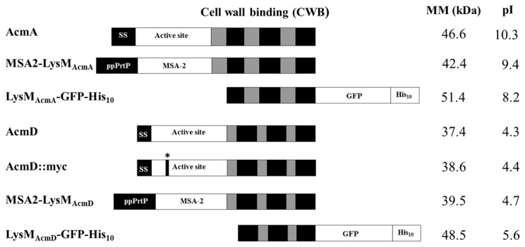
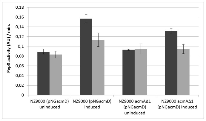
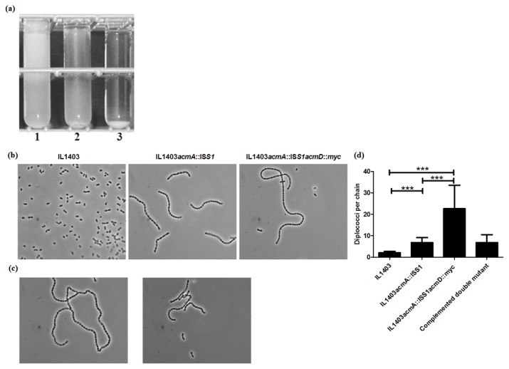
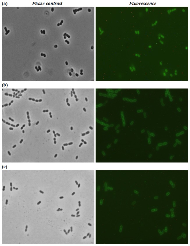
Lactococcus lactis expresses the homologous glucosaminidases AcmB, AcmC, AcmA and AcmD. The latter two have three C-terminal LysM repeats for peptidoglycan binding. AcmD has much shorter intervening sequences separating the LysM repeats and a lower iso-electric point (4.3) than AcmA (10.3). Under standard laboratory conditions AcmD was mainly secreted into the culture supernatant. An L. lactis acmAacmD double mutant formed longer chains than the acmA single mutant, indicating that AcmD contributes to cell separation. This phenotype could be complemented by plasmid-encoded expression of AcmD in the double mutant. No clear difference in cellular lysis and protein secretion was observed between both mutants. Nevertheless, overexpression of AcmD resulted in increased autolysis when AcmA was present (as in the wild type strain) or when AcmA was added to the culture medium of an AcmA-minus strain. Possibly, AcmD is mainly active within the cell wall, at places where proper conditions are present for its binding and catalytic activity. Various fusion proteins carrying either the three LysM repeats of AcmA or AcmD were used to study and compare their cell wall binding characteristics. Whereas binding of the LysM domain of AcmA took place at pHs ranging from 4 to 8, LysM domain of AcmD seems to bind strongest at pH 4.
Conflict of interest statement
Figures
Similar articles
-
AcmA of Lactococcus lactis is an N-acetylglucosaminidase with an optimal number of LysM domains for proper functioning.FEBS J. 2005 Jun;272(11):2854-68. doi: 10.1111/j.1742-4658.2005.04706.x. FEBS J. 2005. PMID: 15943817
-
Autolysis of Lactococcus lactis is increased upon D-alanine depletion of peptidoglycan and lipoteichoic acids.J Bacteriol. 2005 Jan;187(1):114-24. doi: 10.1128/JB.187.1.114-124.2005. J Bacteriol. 2005. PMID: 15601695 Free PMC article.
-
Reduced lysis upon growth of Lactococcus lactis on galactose is a consequence of decreased binding of the autolysin AcmA.Appl Environ Microbiol. 2008 Aug;74(15):4671-9. doi: 10.1128/AEM.00103-08. Epub 2008 Jun 6. Appl Environ Microbiol. 2008. PMID: 18539791 Free PMC article.
-
Molecular cloning and nucleotide sequence of the gene encoding the major peptidoglycan hydrolase of Lactococcus lactis, a muramidase needed for cell separation.J Bacteriol. 1995 Mar;177(6):1554-63. doi: 10.1128/jb.177.6.1554-1563.1995. J Bacteriol. 1995. PMID: 7883712 Free PMC article.
-
The elongation of ovococci.Microb Drug Resist. 2014 Jun;20(3):215-21. doi: 10.1089/mdr.2014.0032. Epub 2014 Apr 28. Microb Drug Resist. 2014. PMID: 24773288 Free PMC article. Review.
Cited by
-
Transcriptional Landscape of a bla KPC-2 Plasmid and Response to Imipenem Exposure in Escherichia coli TOP10.Front Microbiol. 2018 Dec 3;9:2929. doi: 10.3389/fmicb.2018.02929. eCollection 2018. Front Microbiol. 2018. PMID: 30559731 Free PMC article.
-
The protein regulator ArgR and the sRNA derived from the 3'-UTR region of its gene, ArgX, both regulate the arginine deiminase pathway in Lactococcus lactis.PLoS One. 2019 Jun 20;14(6):e0218508. doi: 10.1371/journal.pone.0218508. eCollection 2019. PLoS One. 2019. PMID: 31220124 Free PMC article.
-
A Lactococcus lactis expression vector set with multiple affinity tags to facilitate isolation and direct labeling of heterologous secreted proteins.Appl Microbiol Biotechnol. 2017 Nov;101(22):8139-8149. doi: 10.1007/s00253-017-8524-x. Epub 2017 Oct 2. Appl Microbiol Biotechnol. 2017. PMID: 28971274 Free PMC article.
-
Lantibiotic immunity: inhibition of nisin mediated pore formation by NisI.PLoS One. 2014 Jul 11;9(7):e102246. doi: 10.1371/journal.pone.0102246. eCollection 2014. PLoS One. 2014. PMID: 25014359 Free PMC article.
-
Expression of prophage-encoded endolysins contributes to autolysis of Lactococcus lactis.Appl Microbiol Biotechnol. 2017 Feb;101(3):1099-1110. doi: 10.1007/s00253-016-7822-z. Epub 2016 Sep 22. Appl Microbiol Biotechnol. 2017. PMID: 27660179 Free PMC article.
References
-
- Bateman A, Bycroft M (2000) The structure of a LysM domain from E. coli membrane-bound lytic murein transglycosylase D (MltD). J Mol Biol 299: 1113–1119. - PubMed
-
- Bolotin A, Quinquis B, Ehrlich SD, Sorokin A (2012) Complete genome sequence of Lactococcus lactis subsp. cremoris A76. J Bacteriol 194(5): 1241–1242. doi:10.1128/JB.06629-11. PubMed: 22328746. - DOI - PMC - PubMed
-
- Bolotin A, Wincker P, Mauger S, Jaillon O, Malarme K et al. (2001) The complete genome sequence of the lactic acid bacterium Lactococcus lactis ssp. lactis IL1403. Genome Res 11: 731–753. doi:10.1101/gr.GR -1697R PubMed: 11337471. - DOI - PMC - PubMed
-
- Bosma T, Kanninga R, Neef J, Audouy SAL, van Roosmalen ML et al. (2006) Novel surface display system for proteins on non-genetically modified Gram-positive bacteria. Appl Environ Microbiol 72: 880–889. doi:10.1128/AEM.72.1.880-889.2006. PubMed: 16391130. - DOI - PMC - PubMed
Publication types
MeSH terms
Substances
LinkOut - more resources
Full Text Sources
Other Literature Sources
Molecular Biology Databases
