. 2013 Aug 12;8(8):e73621.
doi: 10.1371/journal.pone.0073621. eCollection 2013.

Adhesion to carbon nanotube conductive scaffolds forces action-potential appearance in immature rat spinal neurons

Affiliations

Adhesion to carbon nanotube conductive scaffolds forces action-potential appearance in immature rat spinal neurons

Alessandra Fabbro et al. PLoS One. .

Abstract

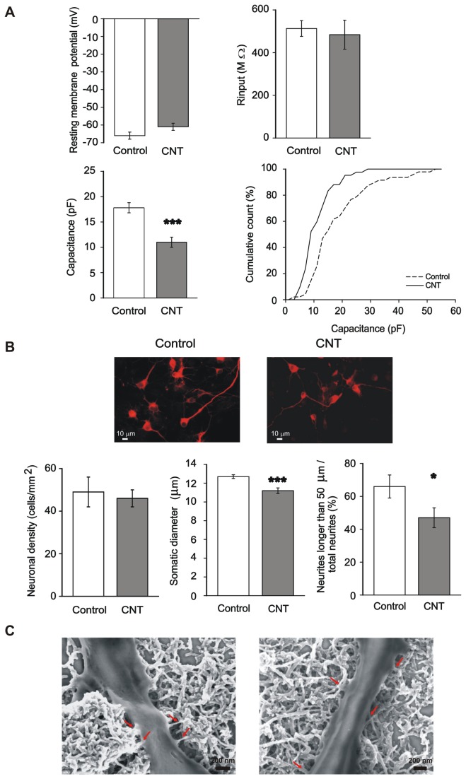
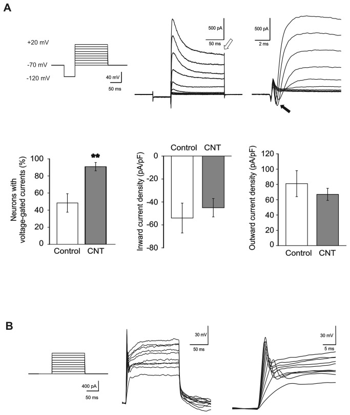
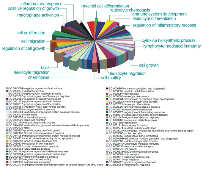
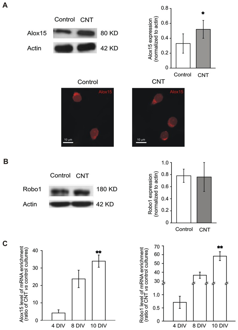
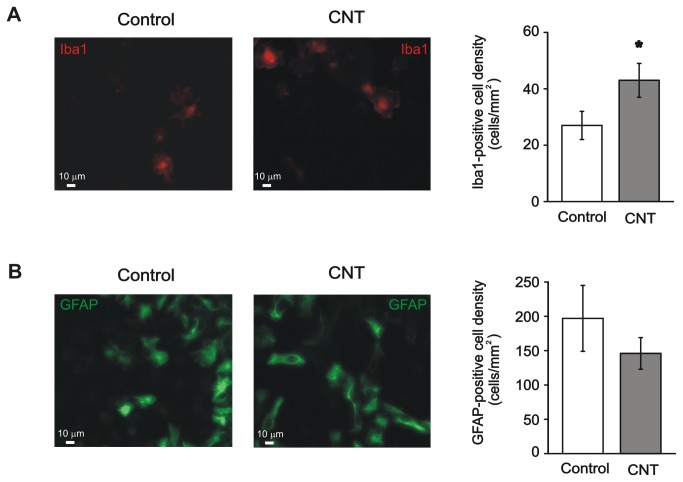
In the last decade, carbon nanotube growth substrates have been used to investigate neurons and neuronal networks formation in vitro when guided by artificial nano-scaled cues. Besides, nanotube-based interfaces are being developed, such as prosthesis for monitoring brain activity. We recently described how carbon nanotube substrates alter the electrophysiological and synaptic responses of hippocampal neurons in culture. This observation highlighted the exceptional ability of this material in interfering with nerve tissue growth. Here we test the hypothesis that carbon nanotube scaffolds promote the development of immature neurons isolated from the neonatal rat spinal cord, and maintained in vitro. To address this issue we performed electrophysiological studies associated to gene expression analysis. Our results indicate that spinal neurons plated on electro-conductive carbon nanotubes show a facilitated development. Spinal neurons anticipate the expression of functional markers of maturation, such as the generation of voltage dependent currents or action potentials. These changes are accompanied by a selective modulation of gene expression, involving neuronal and non-neuronal components. Our microarray experiments suggest that carbon nanotube platforms trigger reparative activities involving microglia, in the absence of reactive gliosis. Hence, future tissue scaffolds blended with conductive nanotubes may be exploited to promote cell differentiation and reparative pathways in neural regeneration strategies.


Conflict of interest statement

Competing Interests: The authors have declared that no competing interests exist.

Figures

Figure 1
Figure 1. Carbon nanotubes substrate selectively affects membrane properties and neurite extension of spinal neurons.
(A) Spinal neurons grown on MWCNT show similar resting potential and membrane resistance (Rinput) with respect to control neurons (top), while membrane capacitance, indicative of total cell membrane extension, is significantly lower in CNT neurons (bottom left, ***: P<0.001; bottom right, cumulative distribution, KS test: P<0.01). (B) Top, typical examples of spinal cord neurons cultured in control condition or on MWCNTs, labeled with the antibody against the neuronal marker β-tubulin III to visualize neuronal morphology. Bottom, despite a similar neuronal density (left), CNT cultures showed a slightly, but significantly lower somatic neuronal diameter compared to control cultures (middle; ***: P<0.001), together with a lower number of long neurites (right; *: P<0.05). These findings are in agreement with the lower membrane capacitance values found in CNT neurons. (C) SEM images of neurites from spinal neurons grown on a MWCNT layer, showing the numerous and very tight contacts between MWCNTs and neuronal membranes (red arrows).
Figure 2
Figure 2. MWCNTs boost the functional maturation of spinal neurons.
(A) Top left, voltage-clamp stimulation protocol to test the presence of voltage-dependent currents. Top middle (and right, in extended time scale), typical recordings from a spinal neuron displaying voltage-dependent currents. Note the presence of both outward (open arrow, middle panel; K+) and inward (filled arrow, right panel; Na+) voltage-dependent currents. The fraction of neurons displaying voltage-dependent currents is considerably higher in CNT neurons with respect to controls (bottom left; **: P<0.01), while current density is similar for inward and outward currents in both culturing conditions (bottom middle and right). (B) Left, current-clamp stimulation protocol to test the neuronal ability to generate action potentials. Middle (and right, in extended time scale), example of action potentials generated by a spinal neuron.
Figure 3
Figure 3. Gene Ontology enrichment analysis showing gene categories whose expression is modulated by MWCNTs.
See Table S2 for statistics.
Figure 4
Figure 4. The expression of Alox15 and Robo1 is increased by MWCNT substrate.
(A) Top left, Western blot analysis of the Alox15 protein (migrated at approximately 80 KD) isolated from control (left lane) and CNT (right lane) spinal cultures, showing the strong increase in Alox15 expression in CNT cultures. Top right, quantification of the Alox15 protein level normalized against actin. Bottom, confocal images of control and CNT neurons, stained using the anti-Alox15 antibody. Note the punctate appearance of strongly Alox15-immunopositive cytoplasmatic structures in both culturing conditions. (B) Left, Western blot analysis of the Robo1 protein (migrated at approximately 180 KD) from control (left lane) and CNT (right lane) cultures. Robo1 expression level is unaffected by the MWCNT substrate (right). (C) Time course of Alox15 (left) and Robo1 (right) expression levels by transcript-specific real time PCR in CNT cultures normalized to control. The expression of both proteins is progressively upregulated in CNT cultures during in vitro development.
Figure 5
Figure 5. MWCNTs impact on the density of non-neuronal cells in spinal cord cultures.
(A) Left, images of control and CNT spinal cultures immunostained for the microglia/macrophage marker Iba1. Right, the density of Iba1-positive cells is increased in CNT cultures (*: P<0.05). (B) Left, images of control and CNT spinal cultures immunostained for the glial marker GFAP. Right, the density of GFAP-positive cells is similar in control and CNT cultures.

References

    1. Place ES, Evans ND, Stevens MM (2009) Complexity in biomaterials for tissue engineering. Nat Mater 8: 457-470. doi:10.1038/nmat2441. PubMed: 19458646. - DOI - PubMed
    1. Dvir T, Timko BP, Kohane DS, Langer R (2011) Nanotechnological strategies for engineering complex tissues. Nat Nanotechnol 6: 13-22. doi:10.1038/nnano.2010.246. PubMed: 21151110. - DOI - PMC - PubMed
    1. Geiger B, Spatz JP, Bershadsky AD (2009) Environmental sensing through focal adhesions. Nat Rev Mol Cell Biol 10: 21-33. doi:10.1038/nrm2593. PubMed: 19197329. - DOI - PubMed
    1. Brunetti V, Maiorano G, Rizzello L, Sorce B, Sabella S et al. (2010) Neurons sense nanoscale roughness with nanometer sensitivity. Proc Natl Acad Sci U S A 107: 6264-6269. doi:10.1073/pnas.0914456107. PubMed: 20308580. - DOI - PMC - PubMed
    1. Atala A (2009) Engineering organs. Curr Opin Biotechnol 20: 575-592. doi:10.1016/j.copbio.2009.10.003. PubMed: 19896823. - DOI - PubMed

Publication types

Substances