. 2012 Aug 1:1:135.
doi: 10.1038/bonekey.2012.135. eCollection 2012.

Mechanisms of multiple myeloma bone disease

Affiliations

Mechanisms of multiple myeloma bone disease

Deborah L Galson et al. Bonekey Rep. .

Abstract

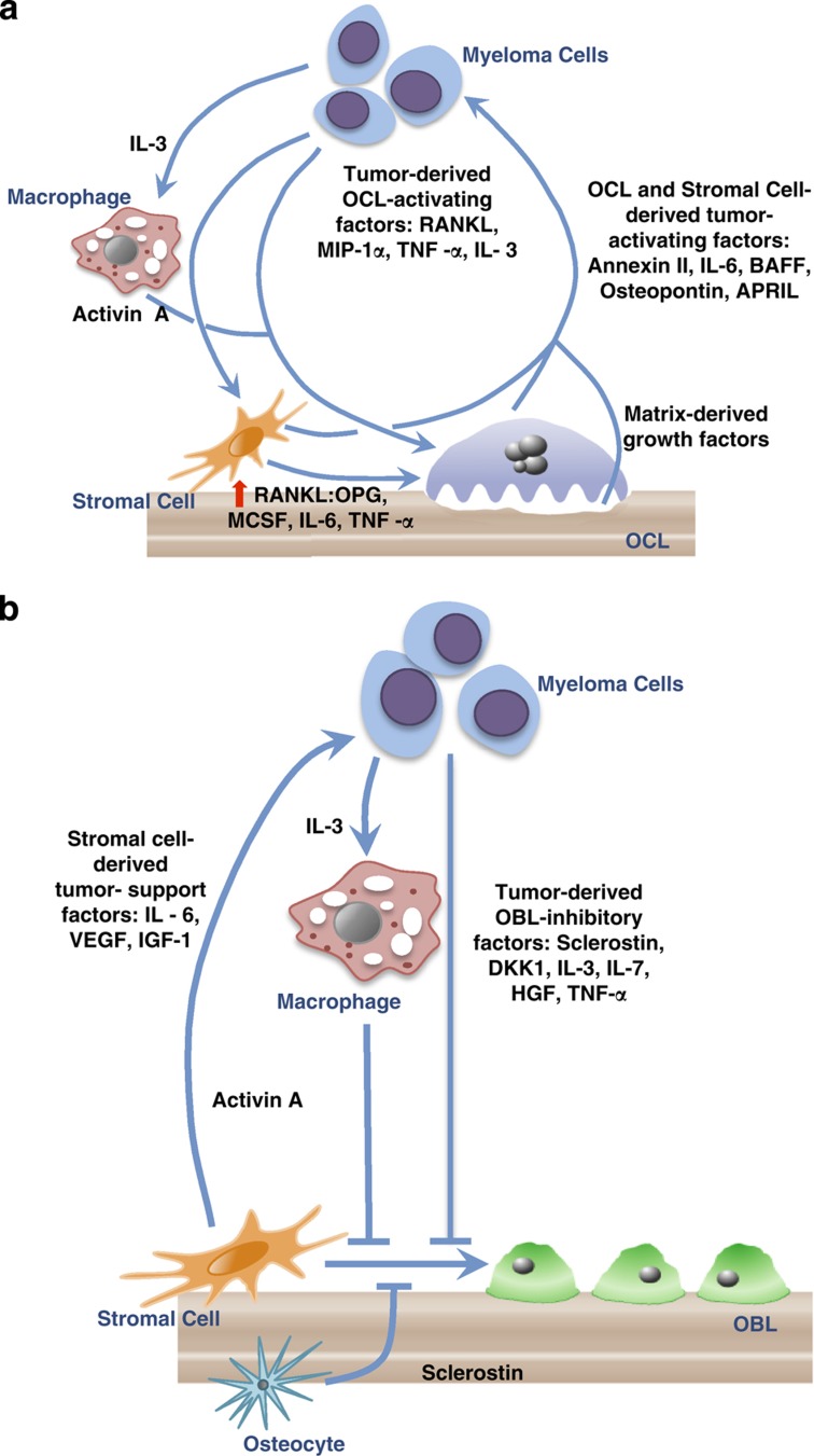
Multiple myeloma is the second most common hematological malignancy and the most frequent cancer to involve the skeleton. Multiple myeloma bone disease (MMBD) is characterized by abnormal bone remodeling with dysfunction of both bone resorption and bone formation, and thus can be used as a paradigm for other inflammatory bone diseases, and the regulation of osteoclasts and osteoblasts in malignancy. Studies of MMBD have identified novel regulators that increase osteoclastogenesis and osteoclast function, repress osteoblast differentiation, increase angiogenesis, or permanently alter stromal cells. This review will discuss the current understanding of mechanisms of osteoclast and osteoblast regulation in MMBD, and therapeutic approaches currently in use and under development that target mediators of bone destruction and blockade of bone formation for myeloma patients, including new anabolic therapies.


Conflict of interest statement

DLG and RS have no conflicts of interest. GDR is a member of the Amgen Advisory Board and is also the local PI for the Denosumab Trial in Myeloma.

Figures

Figure 1
Figure 1
Mechanisms of myeloma bone disease: increased osteoclast (OCL) activity and suppressed osteoblast (OBL) formation in myeloma result in tumor growth and bone destruction. In myeloma bone disease, osteoclastogenesis is favored and osteoblastogenesis is inhibited. (a) Increased OCL activity in myeloma. Multiple factors produced by myeloma cells increase OCL activity. Myeloma cells produce OCL-activating factors (OAFs) that directly increase OCL activity and stimulate marrow stromal cells and T cells to increase production of OAFs and decrease production of OCL inhibitory factors. OAFs produced by myeloma cells include RANKL, MIP-1α, IL-3 and TNF-α. In addition, IL-3 activates tumor-associated macrophages to produce activin A, further increasing OCL activity. Myeloma cells also induce marrow stromal cells production of OAFS, such as RANKL, macrophage colony-stimulating factor (MCSF), IL-6 and TNF-α, and decrease expression of OPG, which enhances OCL formation. Amplifying this process, OCLs and stromal cells secrete soluble factors, such as IL-6, annexin II, osteopontin, BAFF and APRIL, which further stimulate tumor growth. Finally, the enhanced bone destructive process releases growth factors (TGFβ, insulin-like growth factors (IGFs), fibroblast growth factors (FGFs), platelet-derived growth factors (PDGFs), bone morphogenetic proteins (BMPs)) from the bone matrix that increase the growth of myeloma cells, exacerbating the osteolytic process. This results in a 'vicious cycle' of bone destruction. (b) OBL suppression in myeloma. Suppression of OBL differentiation by tumor-derived OBL-inhibitory factors, such as sclerostin, DKK1, IL-3, IL-7, HGF and TNF-α, also has an important role in tumor growth, as mature OBL inhibit myeloma cell growth. In addition, IL-3, secreted by myeloma cells, stimulates release of activin A from macrophages in the bone marrow microenvironment to inhibit osteoblast formation. Myeloma cells also induce cells in the bone microenvironment to increase production of OBL suppressors. Osteocyte production of sclerostin and marrow stromal cell production of TNF-α are examples. TGFβ released from the bone matrix by the enhanced OCL activity in MMBD also inhibits OBL differentiation. Myeloma cells induce changes in marrow stromal cells that increase production of factors that support myeloma cells, such as IL-6, VEGF and IGF-1, in part via adhesive interactions through VCAM-1 on marrow stromal cells and α4β1 on myeloma cells. Finally, bidirectional signaling between ephrin B2 in OCLs and its receptor, EphB4 in BMSC and OBL (not illustrated) negatively control osteoclast formation and promote osteoblast differentiation. Both ephrin B2 and EphB4 are decreased in myeloma, and enhancing ephrinB2–EphB4 signaling to decrease osteoclast function is a possible therapeutic target for MMBD.

References

    1. Diamond T, Levy S, Day P, Barbagallo S, Manoharan A, Kwan YK. Biochemical, histomorphometric and densitometric changes in patients with multiple myeloma: effects of glucocorticoid therapy and disease activity. Br J Haematol 1997;97:641–648. - PubMed
    1. Giuliani N, Rizzoli V, Roodman GD. Multiple myeloma bone disease: pathophysiology of osteoblast inhibition. Blood 2006;108:3992–3996. - PubMed
    1. Mariotto A, Gigli A, Capocaccia R, Tavilla A, Clegg L, Depry M et al.. Complete and limited duration cancer prevalence estimates. SEER Cancer Statistics Review, Bethesda, MD, 2002, pp 19.
    1. Roodman GD. Pathogenesis of myeloma bone disease. Blood Cells Mol Dis 2004;32:290–292. - PubMed
    1. Terpos E, Berenson J, Cook RJ, Lipton A, Coleman RE. Prognostic variables for survival and skeletal complications in patients with multiple myeloma osteolytic bone disease. Leukemia 2010;24:1043–1049. - PubMed

LinkOut - more resources