Review
. 2013 Dec;140(4):391-8.
doi: 10.1111/imm.12162.

The mTOR pathway and integrating immune regulation

Affiliations
Review

The mTOR pathway and integrating immune regulation

Stephen P Cobbold. Immunology. 2013 Dec.

Abstract

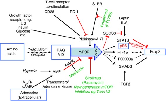
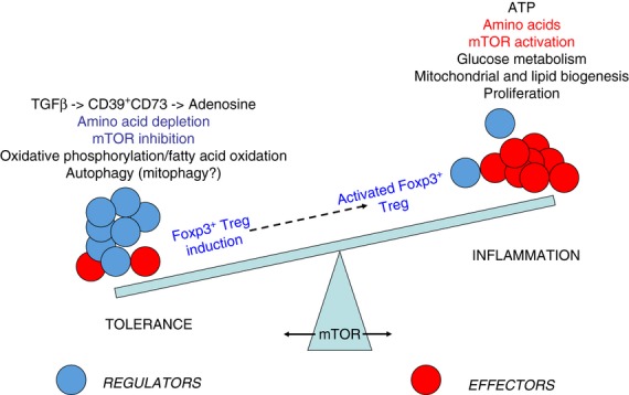
The mammalian target of rapamycin (mTOR) pathway is an important integrator of nutrient-sensing signals in all mammalian cells, and acts to coordinate the cell proliferation with the availability of nutrients such as glucose, amino acids and energy (oxygen and ATP). A large part of the immune response depends on the proliferation and clonal expansion of antigen-specific T cells, which depends on mTOR activation, and the pharmacological inhibition of this pathway by rapamycin is therefore potently immunosuppressive. It is only recently, however, that we have started to understand the more subtle details of how the mTOR pathway is involved in controlling the differentiation of effector versus memory CD8(+) T cells and the decision to generate different CD4(+) helper T-cell subsets. In particular, this review will focus on how nutrient sensing via mTOR controls the expression of the master transcription factor for regulatory T cells in order to maintain the balance between tolerance and inflammation.

Keywords: FOXP3; metabolism; nutrient sensing; regulatory T cells.


Figures

Figure 1
Figure 1
A mammalian target of rapamycin (mTOR) -centric view of nutrient sensing for the induction of forkhead box P3 (FOXP3). The mTOR pathway in T cells integrates antigen receptor signalling through the T-cell receptor and co-stimulatory molecules such as CD28 and programmed death 1 (PD-1) with a range of nutrient-sensing and growth factor signals. The main nutrient sources that are sensed via mTOR are the essential amino acids, glucose and adenosine, while relevant growth factors include insulin and the interleukin-2 family of cytokines. The shingomyelin phosphate receptor (S1PR), which controls lymphocyte exit from the lymph nodes, also acts via this pathway. The availability of amino acids is detected via a unique pathway that involves the ragulator complex and the four Ras-like GTPases (RAGs A–D), activation of which are required before mTOR can form the TORC1 complex and respond to any of the other signals. When the T-cell receptor is stimulated and there are both sufficient nutrients and growth factors available, mTOR is activated, which in turn inhibits FOXP3 expression, so that the differentiation of effector T cells is promoted. When nutrient or growth factor availability is restricted, the resulting mTOR inhibition allows FOXP3 induction and the development of FOXP3+ regulatory T cells. Although the exact mechanism linking mTOR inhibition to FOXP3 expression is not well defined, a number of different pathways have been implicated, as shown. These include direct effects on FOXP3 transcription dependent on hypoxia inducible factor 1α (HIF1α) and forkhead box O3a (FOXO3a), the regulation of FOXP3 mRNA translation by phosphorylation of the ribosomal protein S6 (pS6), and indirect effects on the regulation of FOXP3 expression in response to cytokines such as transforming growth factor-β (TGF-β)and interleukin-6 (IL-6) signalling through SMAD3 or signal transducer and activator of transcription 3 (STAT3), respectively. Positive signalling is indicated by black arrows, while inhibitory signals are indicated by blocked red lines. Broken lines indicate where the details of signalling are still poorly defined or unclear. Approved drugs that are available to manipulate this system in vivo are shown in green.
Figure 2
Figure 2
Transforming growth factor-β (TGF-β) regulates the production of extracellular adenosine. Extracellular ATP released from infections and necrotic cell death is potently inflammatory. Regulatory T (Treg) cells constitutively express the two ectoenzymes CD39 and CD73 that can convert ATP, via AMP into adenosine, thereby converting an inflammatory stimulus into an anti-inflammatory one. TGF-β is, however, able to induce the expression of both CD39 and CD73 on the majority of activated T cells, as well as on other cell types such as macrophages and dendritic cells, thereby providing a mechanism to dramatically amplify the anti-inflammatory action of these two enzymes.
Figure 3
Figure 3
Enzymes that catabolize or use essential amino acids. Each of the amino acids that are considered essential for mammalian cells, because they are unable to synthesize them, are either catabolized (red arrows) or used to synthesize various products (blue arrows) by specific enzymes. Many of these enzymes are up-regulated in dendritic cells in response to inflammation and cytokines (both pro- and anti-inflammatory) or by the action of regulatory T (Treg) cells (highlighted in bold face). For example, arginase 1 (Arg1) and inducible nitric oxide synthase (iNOS/NOS2) can both consume arginine, the availability of which is sensed through the RAG/mammalian target of rapamycin (mTOR) pathway, leading to the inhibition of T-cell proliferation and the promotion of forkhead box P3 (FOXP3) expression (Fig. 1).
Figure 4
Figure 4
Mammalian target of rapamycin (mTOR) at the fulcrum of the immunoregulatory balance. The immune system has to maintain a delicate balance between the inflammation required to protect the body from infection while limiting the potential pathology and risk of autoimmunity. Tolerance is maintained primarily by ensuring that the relative frequency of regulatory T (Treg) cells (blue) is in excess of effector T cells (red), but when an inflammatory response is required, the generation of effector T cells is favoured. The decision to preferentially generate effector T cells rather than Treg cells is dependent on the availability of glucose and essential amino acids, and this activates mTOR, which then coordinates the switch in metabolism from primarily fatty acid oxidation to glucose metabolism. Conversely, the local depletion of essential amino acids from the tolerogenic microenvironment inhibits mTOR and encourages induction of Treg cells over effector cells. Committed induced Treg cells may, however, still be able to respond to mTOR activation by proliferating and/or increasing their suppressive function if they are required to limit the development of any inflammatory pathology.

References

    1. Peter C, Waldmann H, Cobbold SP. mTOR signalling and metabolic regulation of T cell differentiation. Curr Opin Immunol. 2010;22:655–61. - PubMed
    1. Warburg O. On respiratory impairment in cancer cells. Science. 1956;124:269–70. - PubMed
    1. Buttgereit F, Brand MD, Muller M. ConA induced changes in energy metabolism of rat thymocytes. Biosci Rep. 1992;12:381–6. - PubMed
    1. Caro-Maldonado A, Gerriets VA, Rathmell JC. Matched and mismatched metabolic fuels in lymphocyte function. Semin Immunol. 2012;24:405–13. - PMC - PubMed
    1. Wieman HL, Wofford JA, Rathmell JC. Cytokine stimulation promotes glucose uptake via phosphatidylinositol-3 kinase/Akt regulation of Glut1 activity and trafficking. Mol Biol Cell. 2007;18:1437–46. - PMC - PubMed

MeSH terms

Substances