. 2013 Nov 15;456(1):89-98.
doi: 10.1042/BJ20130670.

Histone deacetylase SIRT1 modulates and deacetylates DNA base excision repair enzyme thymine DNA glycosylase

Affiliations

Histone deacetylase SIRT1 modulates and deacetylates DNA base excision repair enzyme thymine DNA glycosylase

Amrita Madabushi et al. Biochem J. .

Abstract

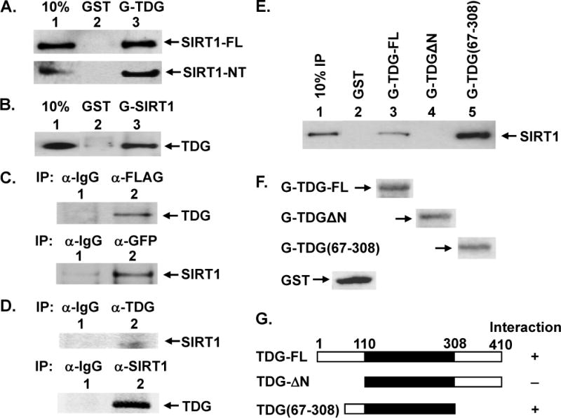
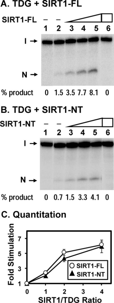
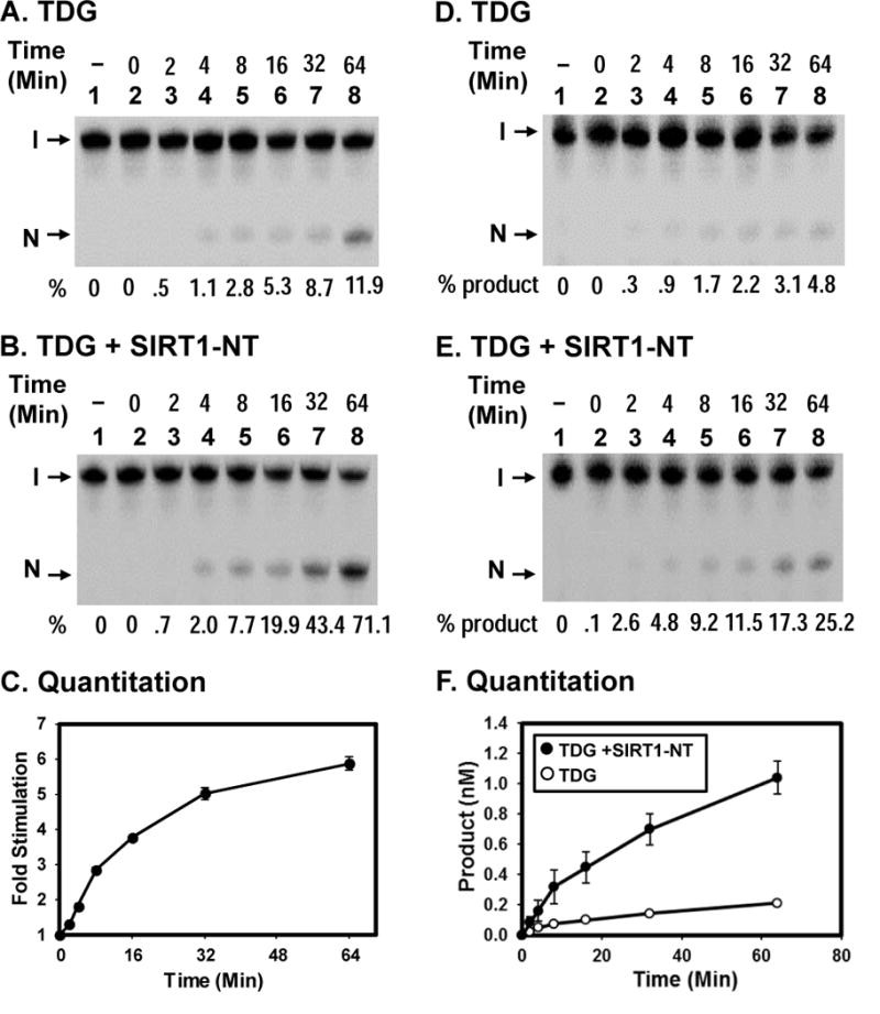
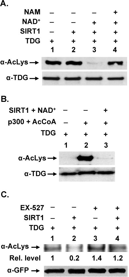
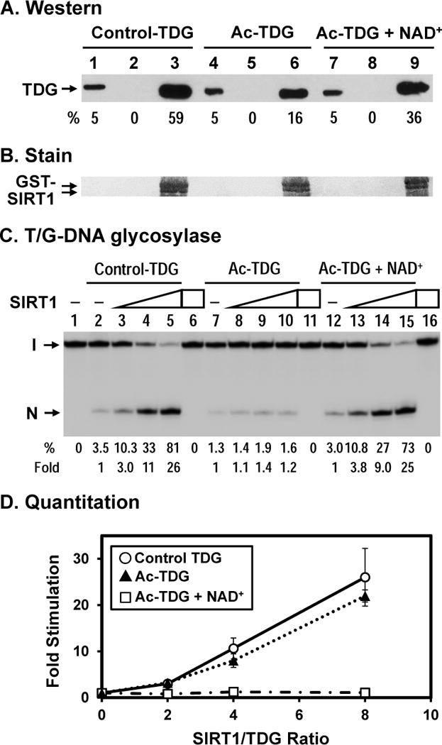
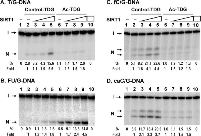
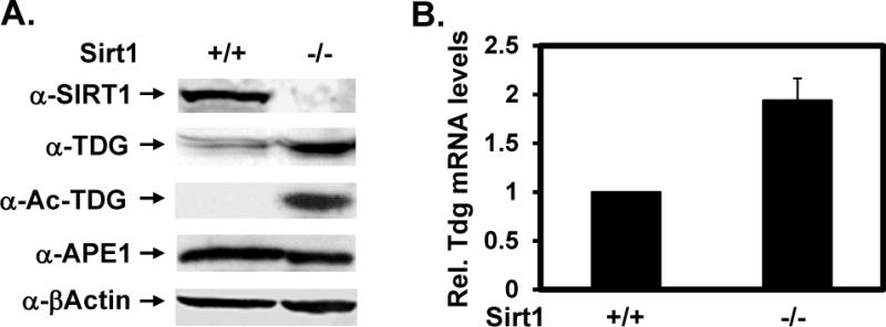
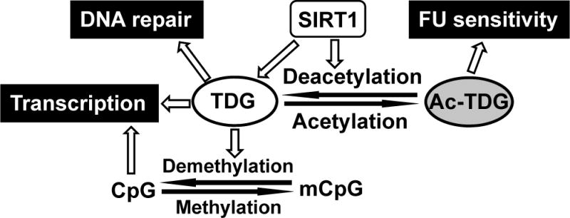
TDG (thymine DNA glycosylase) is an essential multifunctional enzyme involved in DNA base excision repair, DNA demethylation and transcription regulation. TDG is the predominant enzyme that removes thymine from T/G mispair, which arises due to deamination of 5-methyl-cytosine at the CpG dinucleotide, thereby preventing C to T mutations. SIRT1 is a member of class III NAD+-dependent histone/protein deacetylases. In the present study, we demonstrate that SIRT1 interacts with residues 67-110 of hTDG (human TDG). In addition, SIRT1 enhances TDG glycosylase activity and deacetylates acetylated TDG. TDG acetylation weakens its interaction with SIRT1. Although acetylated TDG has reduced glycosylase activity towards T/G, 5-formylcytosine/G and 5-carboxylcytosine/G, it has a stronger activity towards a 5-fluorouracil/G substrate as compared with unmodified TDG. SIRT1 weakly stimulates acetylated hTDG activity towards T/G, 5-formylcytosine/G and 5-carboxylcytosine/G as compared with control hTDG. Sirt1-knockout mouse embryonic fibroblast cells have higher levels of TDG expression and acetylation. The physical and functional interactions between SIRT1 and TDG may mediate DNA repair, gene expression and FU (5-fluorouracil)-mediated cytotoxicity.


Figures

Figure 1
Figure 1. Physical Interaction of TDG with SIRT1
(A) Direct interaction between TDG and SIRT1 was demonstrated by GST pull-down method. GST alone (lane 2) and GST-hTDG (lane 3) were immobilized on glutathione-sepharose and incubated with 100 ng purified FLAG-tagged hSIRT1-FL (upper panel) or 100 ng purified FLAG-tagged hSIRT1-NT (with residues 7–83 deleted) (lower panel) in buffer G (50 mM Tris–HCl, pH 7.4, 150 mM NaCl, and 2 mM EDTA). GST is about 3 fold molar excess of GST-hTDG. The pellets were fractionated on a 10% SDS-polyacrylamide gel followed by Western blot analysis with the FLAG antibody. Lane 1 contains 10 ng (10% of the total input) of FLAG-hSIRT1-FL. (B) Immobilized GST alone (lane 2, in 3–4 fold molar excess) and GST-hSIRT1-NT (lane 3) were incubated with purified His-hTDG. Western blot analysis was performed with the His antibody. Lane 1 contains 10% of the total input of His-hTDG. (C) hTDG and hSIRT1 co-immunoprecipitated in extracts prepared from HEK293T cells expressing FLAG-SIRT1 and GFP-hTDG. Upper panel, immunoprecipation were performed with antibody against FLAG and the Western blot was detected by hTDG antibody (lane 2). Lane 1 is a negative control in which the immunoprecipation was performed with antibody against IgG. Lower panel, immunoprecipation were performed with antibody against GFP and the Western blot was detected by FLAG antibody (lane 2). Lane 1 is a negative control. (D) hTDG and hSIRT1 co-immunoprecipitated in HEK293T extracts. Upper panel, immunoprecipation was performed with antibody against TDG and the Western blot was detected by SIRT1 antibody (lane 2). Lane 1 is a negative control. Lower panel, immunoprecipation was performed with SIRT1 antibody. The Western blot was detected by Cy5-labeled TDG antibody which was prepared according to the manufacturer’s instructions and detected by Typhoon FLA9500 (GE HealthCare). Lane 1 is a negative control. (E) SIRT1 binds to residues 67–110 of TDG. Two TDG deletion constructs were used to determine the region within hTDG involved in binding to SIRT1. FLAG-hSIRT1-FL (100 ng) was incubated with GST-TDG constructs (as labeled) or GST alone immobilized on beads as in (A). (F) The amounts of GST and GST-TDG constructs were determined by Ponceau S stain. The same membrane of (E) was stained by Ponceau S. Due to the large size difference, only the bands of GST-tagged proteins were shown on each lane. As indicated, GST is about 3 fold molar excess of GST-hTDG-FL which is in similar molar amount of GST-TDGΔN and GST-TDG(67–308). (G) Graphic depiction of hTDG constructs and the summary of physical interaction of these constructs with SIRT1. The intact hTDG (TDG-FL) contains 410 amino acid residues and the core catalytic domain is within residues 111–308 (shown in black box). The physical interactions of hTDG and SIRT1 are derived from results of Figure 1E. “+” represents for positive and “−” represents for negative interactions.
Figure 2
Figure 2. Human TDG glycosylase activity can be stimulated by hSIRT1
Human SIRT1-FL (A) and SIRT1-NT (B) purified from HEK293T cells enhance the activities of TDG. Lane 1, T/G-containing DNA substrate. Lane 2, 0.18 nM T/G-containing DNA substrate was incubated with 0.5 nM untreated hTDG Lanes 3–5 are similar to lane 2 but with added 0.5, 1, and 2 nM hSIRT1-FL (or hSIRT1-NT), respectively. Lane 6, T/G-containing DNA substrate was incubated with 2 nM hSIRT1-FL (or hSIRT1-NT) without TDG. The products were then treated with 0.1 M NaOH at 90°C for 30 min and separated on a 14% DNA sequencing gel. The images are pasted together from the same gel. Arrows mark the intact DNA substrate (I) and the cleavage product (N). Percentage (%) of product generated is shown below each lane. (C) Quantitative analyses of the fold of stimulation of hSIRT1-FL (open circles) and hSIRT1-NT (filled triangles) on the TDG glycosylase activity on T/G-containing DNA substrate from three experiments. The error bars reported are the standard deviations of the averages.
Figure 3
Figure 3. Time course of TDG glycosylase reaction under single-turnover and multiple-turnover conditions
(A) Reactions are similar to Fig. 2B (lane 2) with 0.18 nM T/G-DNA and 0.5 nM TDG at various time points as indicated. (B) Reactions are similar to (A) except with 2 nM of SIRT1-NT. Percentage (%) of products generated is shown below each lane. (C) Quantitative analyses of the fold of stimulation of hSIRT1-NT on the TDG glycosylase activity on T/G-containing DNA substrate from three experiments. The error bars reported are the standard deviations of the averages. (D) and (E) Time course of TDG reaction in the absence or presence of hSIRT1-NT, respectively, under multiple-turnover condition. Reactions contained 5 nM T/G-DNA and 0.5 nM TDG in the absence or presence of 2 nM SIRT1-NT. Percentage (%) of products generated is shown below each lane. (F) Plots of product formation vs. time from three experiments as in (D) (open circles) and in (E) (closed circles). The error bars reported are the standard deviations of the averages.
Figure 4
Figure 4. SIRT1 can deacetylate TDG
(A) In vivo acetylated hTDG is a substrate for SIRT1. His-hTDG and p300 plasmids were transiently transfected into HEK293T cells. Acetylated TDG was purified and incubated with SIRT1-FL in the absence or presence of its cofactor NAD+ or its inhibitor nicotinamide (NAM). Western blotting was performed with antibody against acetyl-lysine (α-AcLys) or TDG (α-TDG) to evaluate TDG acetylation. (B) In vitro acetylated hTDG is a substrate for SIRT1. Purified recombinant hTDG protein was incubated with p300 acetyltransferase and acetyl CoA (AcCoA) and then treated with SIRT1-FL in the presence of NAD+. Western blotting was performed with antibody against acetyl-lysine or TDG to evaluate TDG acetylation. (C) SIRT1 controls the in vivo acetylation level of TDG. GFP-hTDG was transiently transfected into HEK293T cells in the absence or presence of SIRT1-FL plasmid. SIRT1 specific inhibitor EX-527 was added 6 hrs before cell harvest. Equal amounts of cell extracts were subjected to immunoprecipitation by anti-GFP. Western blotting was performed with α-acetyl lysine antibody (upper panel) or GFP antibody (lower panel). Relative acetylation level is shown below each lane on upper panel.
Figure 5
Figure 5. Effect of acetylation on TDG interaction with SIRT1
(A) Acetylated TDG has a reduced physical interaction with hSIRT1. hTDG was acetylated in vitro by p300 (lanes 4–9) and control TDG was treated similarly but without acetyl-CoA (lanes 1–3). GST alone (lanes 2, 5, and 8) and GST-hSIRT1 (lanes 3, 6, and 9) were immobilized on glutathione-sepharose and incubated with 100 ng TDG in buffer S (50 mM Tris–HCl, pH 8.0, 150 mM NaCl, and 4 mM MgCl2). Lane 1, 10 ng His-tagged TDG (5% of input), lane 2, unacetylated control TDG bound to GST alone, and lane 3, control TDG bound to GST-SIRT1-FL immobilized on beads. Lanes 4–6 are similar to lanes 1–3 except using in vitro acetylated TDG. Lanes 7–9 are similar to lanes 4–6 except adding 3 mM NAD+ in the incubation buffer. Western blot was detected by TDG primary antibody made in our laboratory and anti-rabbit IgG DyLight 800 (Thermo Scientific) secondary antibody. The image was detected by Typhoon FLA9500 and quantitated by ImageQuant software (GE Healthcare). Percentage (%) of input TDG (lanes 1, 4, and 7) and precipitated TDG as compared to input TDG and adjusted with GST-SIRT1 amounts on membrane in (B) are shown below each lane. (B) The GST-SIRT1 immobilized on membrane of (A) was stained by Ponceau S. As indicated, the amounts of GST-SIRT1 are about the same. (C) SIRT1 has a reduced stimulation on acetylated TDG with T/G-DNA. Control hTDG and acetylated TDG were the same used in (A). 0.18 nM T/G-DNA was incubated with 10 nM TDG in reaction buffer (50 mM Tris-HCl, pH 8.0, 1 mM DTT, 50 μg/ml BSA, 37.5 mM NaCl, 0.75 mM KCl, 1 mM MgCl2) in the absence or presence of SIRT1 or 3 mM NAD+. Lanes 1, DNA alone, Lane 2, 0.18 nM T/G-containing DNA substrate was incubated with 10 nM control hTDG Lanes 3–5 are similar to lane 2 but with added 20, 40, and 80 nM hSIRT1-FL, respectively. Lane 6, T/G-DNA was incubated with 80 nM hSIRT1-FL without TDG. Lanes 7–10 are similar to lanes 2–5 except using Ac-TDG. Lanes 12–15 are similar to lanes 7–10 except adding 3 mM NAD+. Percentage (%) of product generated and fold of stimulation of hSIRT1-FL on the hTDG glycosylase activity from three experiments are shown below each lane. (C) Quantitative analyses of the fold of stimulation of hSIRT1-FL on the TDG glycosylase activity on T/G-DNA substrate from three experiments. Open circles, control TDG; closed triangles, Ac-TDG in the absence of NAD+; and open rectangulars, Ac-TDG in the presence of NAD+. The error bars reported are the standard deviations of the averages.
Figure 6
Figure 6. Effect of acetylation on TDG subtract specificity
(A) Human SIRT1-FL enhances the T/G glycosylase activities of TDG and acetylated TDG. hTDG was acetylated in vitro by p300 (lanes 6–9) and control TDG was treated similarly but without acetyl-CoA (lanes 2–5). Lanes 1–5 and 6–9 are similar to lanes 1–5 in Fig. 2A. Lanes 3–5 and 7–9, 0.18 nM T/G-containing DNA substrate was incubated with 0.5 nM hTDG with added 0.5, 1, and 2 nM hSIRT1-FL, respectively. Lane 10, T/G-containing DNA substrate was incubated with 2 nM hSIRT1-FL without TDG. Percentage (%) of product generated and fold of stimulation of hSIRT1-FL on the hTDG glycosylase activity are shown below each lane. (B) Acetylated hTDG has a stronger activity on FU/G mismatches and can be better stimulated by SIRT1 as compared to unmodified TDG. hTDG glycosylase activity was assayed similarly as in (A) except using 0.2 nM TDG with 0.18 nM FU/G substrate in the presence of 0.2, 0.4, and 0.8 nM of SIRT1-FL. (C) and (D), Acetylated hTDG on fC/G and caC/G mismatches, respectively, can be stimulated by SIRT1. hTDG glycosylase activity was assayed similarly as in (A) except using fC/G or caC/G substrate in the presence of 0.5, 1, and 2 nM of SIRT1-NT and the reaction products were heated at 80°C for 12 min to avoid non-enzyme catalyzed strand cleavage at fC and caC. Under this condition, the AP site is not completely processed by β/δ-elimination, thus two cleavage products (labeled as N) were observed: the upper one is derived by β-elimination only and the lower one is produced by β/δ-elimination.
Figure 7
Figure 7. TDG is overexpressed in sirt1 knockout cells
(A) Protein expression levels in wild-type (+/+) and sirt1 knockout (−/−) MEFs. Western blotting was performed with antibodies against SIRT1, TDG, Ac-TDG, APE1, and β-actin in total cell extracts. The TDG protein levels were normalized to the amounts of β-actin. (B) The Tdg mRNA levels in wild-type (+/+) and sirt1 knockout (−/−) MEFs. The mRNA levels were measured by RT-qPCR analysis. The mRNA level of Tdg was calculated relative to that of β-actin as described in Supplementary Materials. The relative Tdg mRNA levels of WT cells over sirt1 KO cells are presented in X-axis. The reactions were carried out in duplicate and data are averaged from three independent experiments.
Figure 8
Figure 8. A model depicting the role of SIRT1 in mediating TDG function in DNA repair, gene expression, and drug cytotoxicity
SIRT1 enhances TDG glycosylase activity, deacetylates TDG, and suppresses TDG gene expression. Acetylated TDG has reduced glycosylase activities toward T/G, fC/G, and caC/G mismatches, but has a stronger activity toward FU/G substrate as compared to unmodified TDG. Thus, we propose that unmodified TDG is responsible for DNA repair of T/G mismatches and DNA demethylation while acetylated TDG promotes FU removal. If the resulting AP sites are not efficiently repaired by BER after FU removal, spontaneous breakage at AP sites leads to the persistence of single-strand DNA lesions. During DNA replication, these DNA nicks can be converted to double-strand breaks. Alternatively, spontaneous breakage at nearby AP sites generates double-strand breaks and thus causes FU cytotoxicity and cell death.

Similar articles

Cited by

References

    1. Krokan HE, Nilsen H, Skorpen F, Otterlei M, Slupphaug G. Base excision repair of DNA in mammalian cells. FEBS Lett. 2000;476:73–77. - PubMed
    1. Cortazar D, Kunz C, Saito Y, Steinacher R, Schar P. The enigmatic thymine DNA glycosylase. DNA Repair (Amst) 2007;6:489–504. - PubMed
    1. Bennett MT, Rodgers MT, Hebert AS, Ruslander LE, Eisele L, Drohat AC. Specificity of human thymine DNA glycosylase depends on N-glycosidic bond stability. J Am Chem Soc. 2006;128:12510–12519. - PMC - PubMed
    1. Hardeland U, Bentele M, Lettieri T, Steinacher R, Jiricny J, Schar P. Thymine DNA glycosylase. Prog Nucleic Acid Res Mol Biol. 2001;68:235–253. - PubMed
    1. Hardeland U, Bentele M, Jiricny J, Schar P. The versatile thymine DNA-glycosylase: a comparative characterization of the human, Drosophila and fission yeast orthologs. Nucleic Acids Res. 2003;31:2261–2271. - PMC - PubMed

Publication types