

Validation of shear wave elastography in skeletal muscle
- PMID: 23953670
- PMCID: PMC3818126
- DOI: 10.1016/j.jbiomech.2013.07.033
Validation of shear wave elastography in skeletal muscle
Abstract
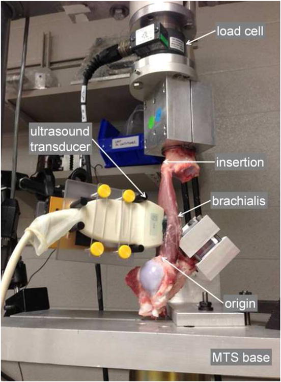
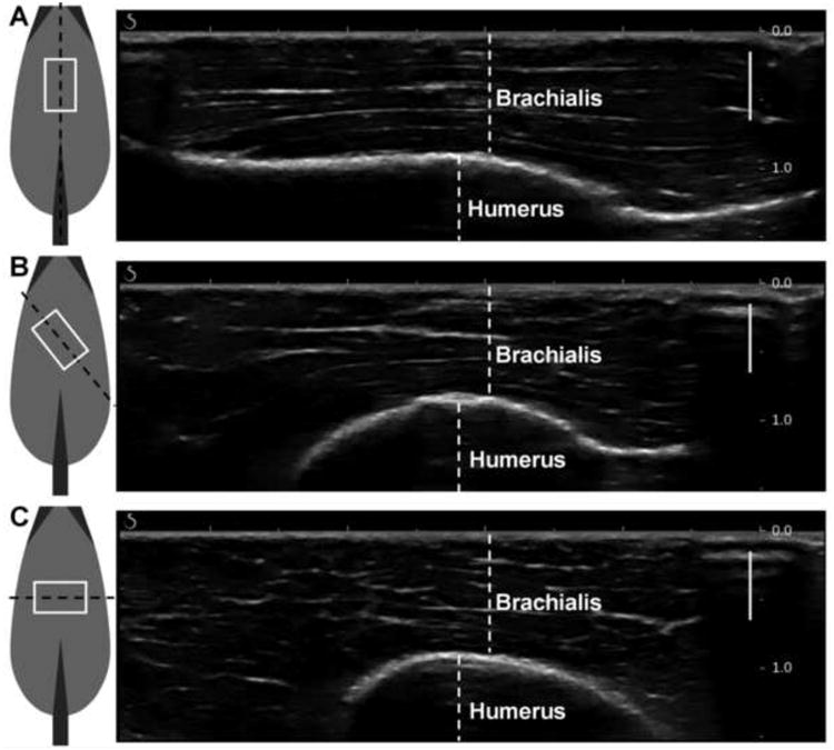
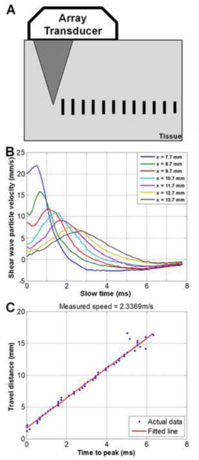
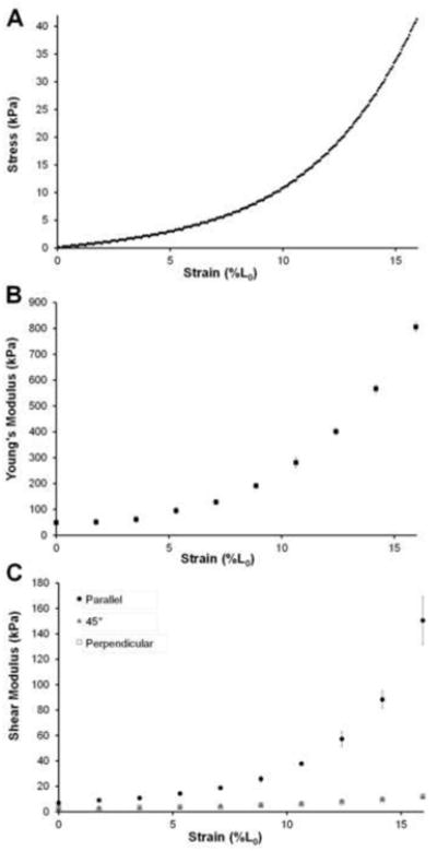
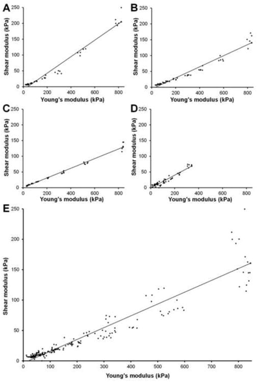
Skeletal muscle is a very dynamic tissue, thus accurate quantification of skeletal muscle stiffness throughout its functional range is crucial to improve the physical functioning and independence following pathology. Shear wave elastography (SWE) is an ultrasound-based technique that characterizes tissue mechanical properties based on the propagation of remotely induced shear waves. The objective of this study is to validate SWE throughout the functional range of motion of skeletal muscle for three ultrasound transducer orientations. We hypothesized that combining traditional materials testing (MTS) techniques with SWE measurements will show increased stiffness measures with increasing tensile load, and will correlate well with each other for trials in which the transducer is parallel to underlying muscle fibers. To evaluate this hypothesis, we monitored the deformation throughout tensile loading of four porcine brachialis whole-muscle tissue specimens, while simultaneously making SWE measurements of the same specimen. We used regression to examine the correlation between Young's modulus from MTS and shear modulus from SWE for each of the transducer orientations. We applied a generalized linear model to account for repeated testing. Model parameters were estimated via generalized estimating equations. The regression coefficient was 0.1944, with a 95% confidence interval of (0.1463-0.2425) for parallel transducer trials. Shear waves did not propagate well for both the 45° and perpendicular transducer orientations. Both parallel SWE and MTS showed increased stiffness with increasing tensile load. This study provides the necessary first step for additional studies that can evaluate the distribution of stiffness throughout muscle.
Keywords: Elastic moduli; Materials testing; Passive stiffness; Shear wave elastography; Ultrasonography.
Copyright © 2013 Elsevier Ltd. All rights reserved.
Conflict of interest statement
Conflict of interest statement: The authors do not have any financial or personal relationships to disclose that could have inappropriately biased this work.
Figures
Similar articles
-
Ultrasound shear wave elastography in assessment of muscle stiffness in patients with Parkinson's disease: a primary observation.Clin Imaging. 2016 Nov-Dec;40(6):1075-1080. doi: 10.1016/j.clinimag.2016.05.008. Epub 2016 May 29. Clin Imaging. 2016. PMID: 27408992
-
In vivo assessment of shear modulus along the fibers of pennate muscle during passive lengthening and contraction using steered ultrasound push beams.J Mech Behav Biomed Mater. 2025 Mar;163:106862. doi: 10.1016/j.jmbbm.2024.106862. Epub 2024 Dec 8. J Mech Behav Biomed Mater. 2025. PMID: 39662288
-
Non-invasive Quantitative Assessment of Muscle Force Based on Ultrasonic Shear Wave Elastography.Ultrasound Med Biol. 2019 Feb;45(2):440-451. doi: 10.1016/j.ultrasmedbio.2018.07.009. Epub 2018 Nov 3. Ultrasound Med Biol. 2019. PMID: 30396600
-
Measuring Shear Wave Velocity in Adult Skeletal Muscle with Ultrasound 2-D Shear Wave Elastography: A Scoping Review.Ultrasound Med Biol. 2023 Jun;49(6):1353-1362. doi: 10.1016/j.ultrasmedbio.2023.02.005. Epub 2023 Mar 21. Ultrasound Med Biol. 2023. PMID: 36958957
-
Using Shear-Wave Elastography to Assess Exercise-Induced Muscle Damage: A Review.Sensors (Basel). 2022 Oct 6;22(19):7574. doi: 10.3390/s22197574. Sensors (Basel). 2022. PMID: 36236672 Free PMC article. Review.
Cited by
-
Acute muscle and joint mechanical responses following a high-intensity stretching protocol.Eur J Appl Physiol. 2016 Aug;116(8):1519-26. doi: 10.1007/s00421-016-3410-2. Epub 2016 Jun 7. Eur J Appl Physiol. 2016. PMID: 27270900 Clinical Trial.
-
Quantifying uniaxial prestress and waveguide effects on dynamic elastography estimates for a cylindrical rod.J Acoust Soc Am. 2023 Dec 1;154(6):3580-3594. doi: 10.1121/10.0022581. J Acoust Soc Am. 2023. PMID: 38038614 Free PMC article.
-
Intra-Rater Reliability of Shear Wave Elastography for the Quantification of Respiratory Muscles in Adolescent Athletes.Sensors (Basel). 2022 Sep 1;22(17):6622. doi: 10.3390/s22176622. Sensors (Basel). 2022. PMID: 36081075 Free PMC article.
-
Mapped Chebyshev pseudo-spectral method for simulating the shear wave propagation in the plane of symmetry of a transversely isotropic viscoelastic medium.Med Biol Eng Comput. 2017 Mar;55(3):389-401. doi: 10.1007/s11517-016-1522-9. Epub 2016 May 25. Med Biol Eng Comput. 2017. PMID: 27221812 Free PMC article.
-
The Association between Tensiomyography and Elastography Stiffness Measurements in Lower Limb Skeletal Muscles.Sensors (Basel). 2022 Feb 5;22(3):1206. doi: 10.3390/s22031206. Sensors (Basel). 2022. PMID: 35161950 Free PMC article.
References
-
- Bensamoun SF, Stevens L, Fleury M, Goubel F, Tho MH. Macroscopic-microscopic characterization of the passive mechanical properties in rat soleus muscle. Journal of Biomechanics. 2006;39:568–578. - PubMed
-
- Bensamoun SF, Wang L, Robert L, Charleux F, Latrive J, Tho MH. Measurement of liver stiffness with two imaging techniques: magnetic resonance elastography and ultrasound elastometry. Journal of Magnetic Resonance Imaging : JMRI. 2008;28:1287–92. - PubMed
-
- Bercoff J, Tanter M, Fink M. Supersonic shear imaging: a new technique for soft tissue elasticity mapping. IEEE Transactions on Ultrasonics, Ferroelectrics, and Frequency Control. 2004;51:396–409. - PubMed
-
- Bizzini M, Mannion AF. Reliability of a new, hand-held device for assessing skeletal muscle stiffness. Clinical Biomechanics. 2003;18:459–61. - PubMed
Publication types
MeSH terms
Grants and funding
LinkOut - more resources
Full Text Sources
Other Literature Sources
Medical
