Review
. 2013 May;33(3):237-47.
doi: 10.1016/j.semnephrol.2013.04.004.

Extracellular potassium homeostasis: insights from hypokalemic periodic paralysis

Affiliations
Review

Extracellular potassium homeostasis: insights from hypokalemic periodic paralysis

Chih-Jen Cheng et al. Semin Nephrol. 2013 May.

Abstract

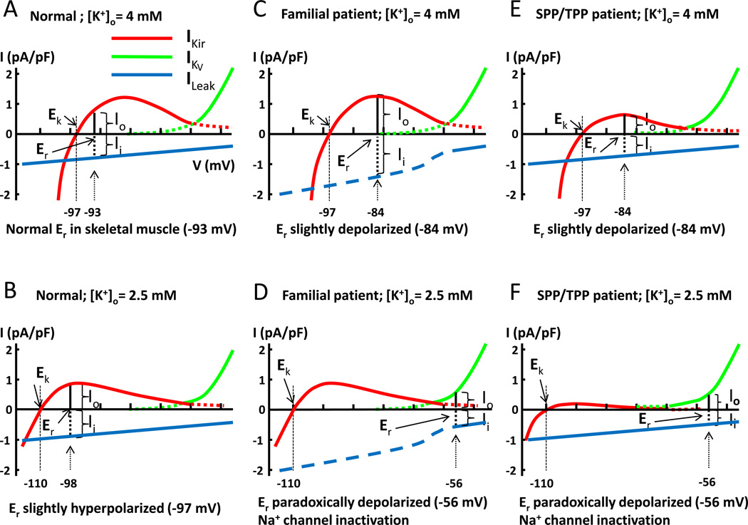
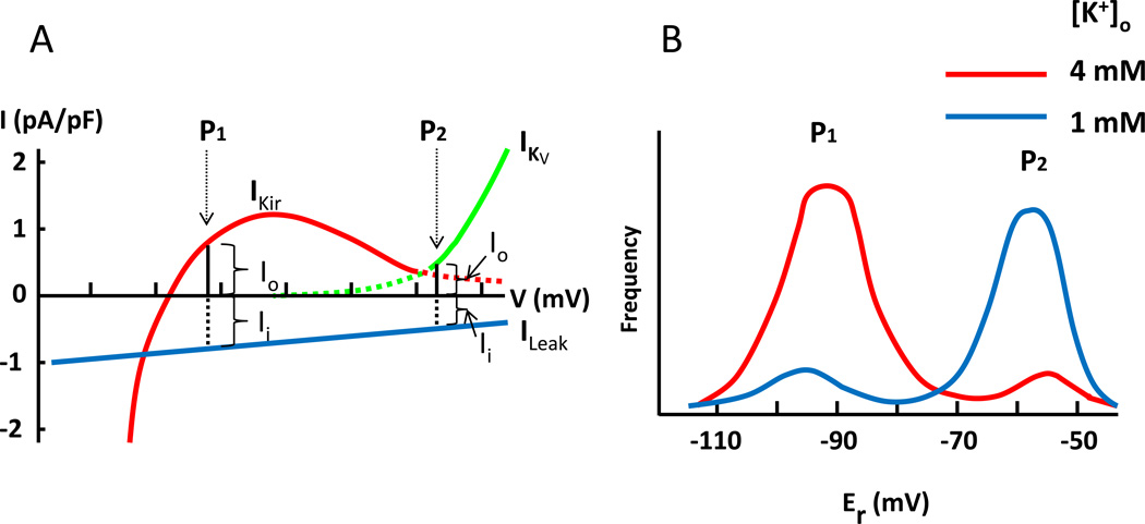
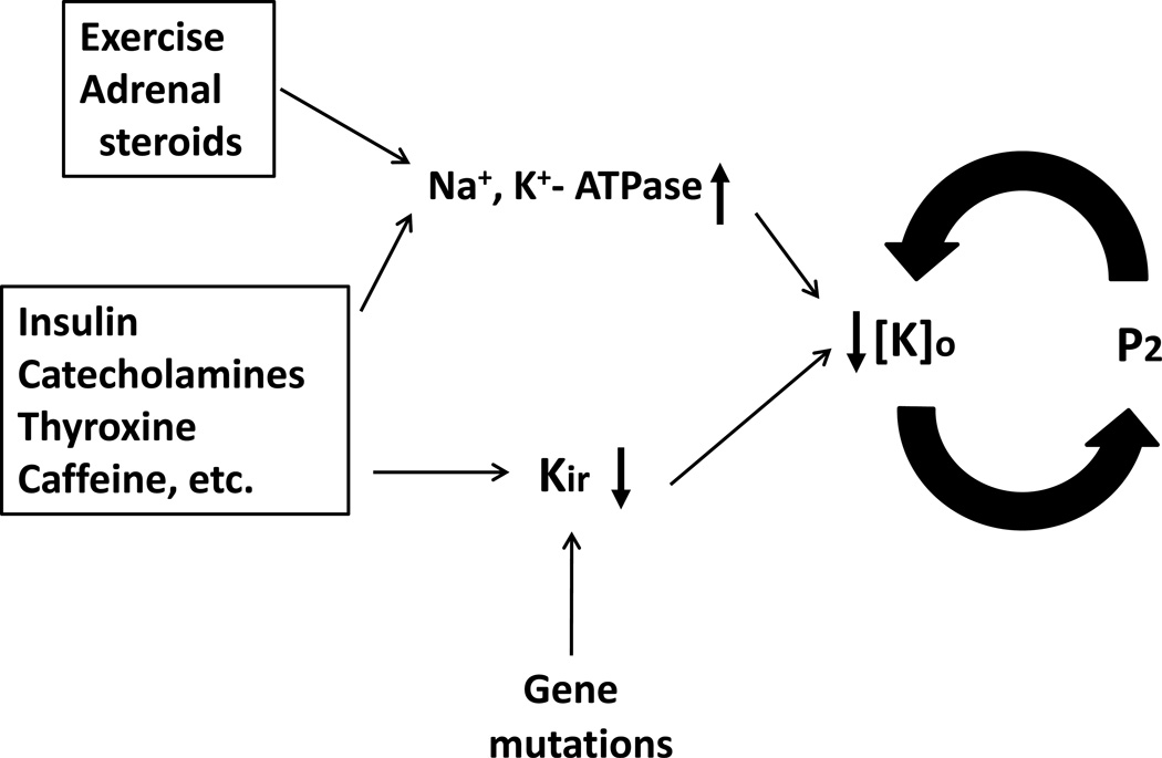
Extracellular potassium makes up only about 2% of the total body's potassium store. The majority of the body potassium is distributed in the intracellular space, of which about 80% is in skeletal muscle. Movement of potassium in and out of skeletal muscle thus plays a pivotal role in extracellular potassium homeostasis. The exchange of potassium between the extracellular space and skeletal muscle is mediated by specific membrane transporters. These include potassium uptake by Na(+), K(+)-adenosine triphosphatase and release by inward-rectifier K(+) channels. These processes are regulated by circulating hormones, peptides, ions, and by physical activity of muscle as well as dietary potassium intake. Pharmaceutical agents, poisons, and disease conditions also affect the exchange and alter extracellular potassium concentration. Here, we review extracellular potassium homeostasis, focusing on factors and conditions that influence the balance of potassium movement in skeletal muscle. Recent findings that mutations of a skeletal muscle-specific inward-rectifier K(+) channel cause hypokalemic periodic paralysis provide interesting insights into the role of skeletal muscle in extracellular potassium homeostasis. These recent findings are reviewed.

Keywords: Hypokalemic periodic paralysis; K(+)-ATPase; Kir; Na(+); hypokalemia-induced paradoxical depolarization; inward-rectifier K(+) channel; skeletal muscle; thyrotoxic periodic paralysis.


Figures

Figure 1
Figure 1
Models for paradoxical depolarization in patients with hypokalemic periodic paralysis. Current-voltage (I-V) relationship curves for inward rectifier K+ channel (Kir, red curve), voltage-gated K+ channel (KV, green curve), and leak current (blue line). The reversal potential of leak current is 0 mV. Please note that aberrant pore current from mutations of Ca2+ and Na+ channels in familial HypoPP patients only exists in hyperpolarized potentials. Thus, the total inward leak current in familial HypoPP patients is not linear, but is increased in hyperpolarized potentials (shown in interrupted blue line). The model is intended for conceptual understanding; numerical value may be slightly different from true in vivo value. Abbreviations: Ek: equilibrium potential for K+; Er: resting membrane potential; Io: outward cation current; Ii: inward cation current; IKir: current of inward rectifier K+ channel; IKV: current of voltage-gated K+ channel; ILeak: inward cation leak current; [K]o: extracellular potassium concentration. See text for further details.
Figure 2
Figure 2
Decreased potassium efflux at paradoxical depolarization (A) and bi-modal distribution of resting membrane potentials (B). (A) Leak current is a function of membrane potential. Inward leak current at the normal hyperpolarized membrane potential (“P1”) is larger than that at the paradoxically depolarized membrane potential (“P2”). Thus, K+ efflux at P1 (mediated by Kir channel) is larger than that at P2 (mediated by KV channel). (B) Because of heterogeneity, muscle fibers develop paradoxical depolarization at different [K+]o. This is reflected by bi-modal distribution of resting membrane potentials (Er) of a population of muscle fibers. Red and blue curve represent the distribution of Er at [K+]o 4 mM and 1 mM, respectively. Note that shown here is the distribution of normal muscle fibers, in which paradoxical depolarization occurs at extreme hypokalemia ([K+]o ~1 mM). In HypoPP, muscles fibers develop paradoxical depolarization at a relatively higher [K+]o, ~2.5 mM. That is, increased inward leak current as produced by mutations of Ca2+ and Na+ channel in familial HypoPP or decreased outward K+ current as caused by mutations of Kir2.6 in TPP and SPP predispose muscle fibers to paradoxical depolarization. See text for further details.
Figure 3
Figure 3
A positive feedback cycle for development of severe hypokalemia via paradoxical depolarization of skeletal muscle membrane potential. Exercise and adrenal steroids stimulate Na+, K+-ATPase. Insulin, catecholamines, thyroid hormones, caffeine, etc can both stimulate Na+, K+-ATPase and inhibit Kir. These effects can lead to hypokalemia. Loss-of-function mutations of Kir predispose to the development of severe hypokalemia through a positive feedback cycle between paradoxical depolarization and hypokalemia. Combined stimulation of Na+, K+-ATPase and inhibition of Kir by endogenous and/or exogenous factors may cause significant hypokalemia to set in motion the positive feedback cycle for paradoxical depolarization without Kir gene mutation. See text for further details.

References

    1. Sejersted OM, Sjøgaard G. Dynamics and consequences of potassium shifts in skeletal muscle and heart during exercise. Physiol Rev. 2000;80:1411–1481. - PubMed
    1. Sjøgaard G, Adams RP, Saltin B. Water and ion shifts in skeletal muscle of humans with intense dynamic knee extension. Am J Physiol. 1985;248:R190–R196. - PubMed
    1. Youn JH, McDonough AA. Recent advances in understanding integrative control of potassium homeostasis. Annu Rev Physiol. 2009;71:381–401. - PMC - PubMed
    1. Skou JC. The influence of some cations on an adenosine triphosphatase from peripheral nerves. Biochim Biophys Acta. 1957;23:394–401. - PubMed
    1. Skou JC. Enzymatic basis for active transport of Na+ and K+ across cell membrane. Physiol Rev. 1965;45:596–617. - PubMed

Publication types

MeSH terms

Substances