. 2013 Aug 14;14(2):171-182.
doi: 10.1016/j.chom.2013.07.010.

Salmonella require the fatty acid regulator PPARδ for the establishment of a metabolic environment essential for long-term persistence

Affiliations

Salmonella require the fatty acid regulator PPARδ for the establishment of a metabolic environment essential for long-term persistence

Nicholas A Eisele et al. Cell Host Microbe. .

Abstract

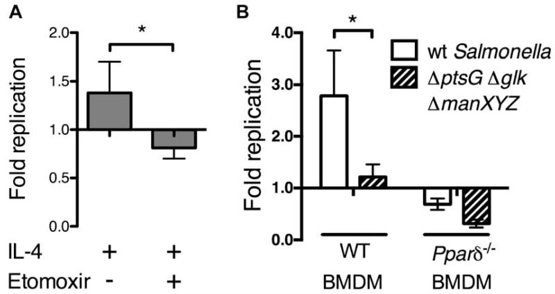
Host-adapted Salmonella strains are responsible for a number of disease manifestations in mammals, including an asymptomatic chronic infection in which bacteria survive within macrophages located in systemic sites. However, the host cell physiology and metabolic requirements supporting bacterial persistence are poorly understood. In a mouse model of long-term infection, we found that S. typhimurium preferentially associates with anti-inflammatory/M2 macrophages at later stages of infection. Further, PPARδ, a eukaryotic transcription factor involved in sustaining fatty acid metabolism, is upregulated in Salmonella-infected macrophages. PPARδ deficiency dramatically inhibits Salmonella replication, which is linked to the metabolic state of macrophages and the level of intracellular glucose available to bacteria. Pharmacological activation of PPARδ increases glucose availability and enhances bacterial replication in macrophages and mice, while Salmonella fail to persist in Pparδ null mice. These data suggest that M2 macrophages represent a unique niche for long-term intracellular bacterial survival and link the PPARδ-regulated metabolic state of the host cell to persistent bacterial infection.


Figures

Figure 1
Figure 1. S. typhimurium resides in anti-inflammatory/M2-like macrophages during chronic infection
(A) Bi-phasic immune response in infected MLNs. Concentrations of cytokines were determined by multiplex analysis at the indicated time points after infection. Each block corresponds to one cytokine value in one mouse. Results from one representative experiment are shown. 1 and 2 indicate the two clusters of cytokines identified based on similar expression profiles along the time course of infection. Cluster 1 contains pro-inflammatory cytokines and cluster 2 shows anti-inflammatory factors. (B) Macrophages from the spleen and MLNs were identified based on CD301 cell surface expression in CD11b+ F4/80+ macrophages. Percentage of CD301+ cells relative to the total number of cells from the corresponding tissue (grey bars, left y axis), and Salmonella colonization level from the same organs (solid black line, right y axis) are shown. Data presented are representative of two independent experiments. (C) Splenocytes were isolated at 30 dpi and CD301+ macrophages were identified based on CD301 expression of CD11b+ F4/80+ Ly6G-cells. Salmonella-positive gates were drawn based on plots from samples stained with an appropriate isotype control antibody. Numbers indicate the percentage of positive cells in each gate. The data presented are representative of three independent experiments. (D) Salmonella-associated mean fluorescence intensity (MFI) associated with CD301+ or CD301 macrophages from the spleen at 30 dpi. Graph shows the means ± SD of five mice and are representative of at least three independent experiments. (E) WT or Il-4raLoxP/LoxP-LysmCre BALB/c mice were infected orogastrically with 109 CFU S. typhimurium SL1344ΔaroA and bacterial loads in the MLNs were quantified 28 dpi. Shown is the geometric mean determined from two independent experiments. All other data are presented as mean ± SD. *p< 0.05, **p< 0.01. Figure 1, related to Figure S1.
Figure 2
Figure 2. Enhanced S. typhimurium replication within IL-4-stimulated cells
(A) Salmonella replication in BMDMs treated as shown. At the indicated time points, cells were lysed and bacterial loads were enumerated. (B) Cell death was determined by measuring LDH release at 26 hpi and is represented as the percentage of total cell death in lysed control cells. (C) Relative proportion of Salmonella-associated cells in the CD301+ and CD301 populations from IL-4-stimulated BMDMs at 2 hpi. (D) Salmonella MFI of CD301+ or CD301 macrophages at 26 hpi. (E) Fold replication of Salmonella in CD301+ and CD301 populations. Data are presented as mean ± SD. *p< 0.05, **p< 0.01. Graphs show the means of triplicate samples and are representative of at least three independent experiments. Figure 2, related to Figure S2.
Figure 3
Figure 3. PPARδ is required for Salmonella replication
(A) Up-regulation of Pparδ mRNA in S. typhimurium-infected BMDMs treated with IL-4. Relative transcript levels for genes encoding Ppar family members are shown. Data were normalized to uninfected samples for each gene. (B) Salmonella 12023 replication in IL-4-stimulatd WT and Pparδ−/− BMDMs. (C) Percentage of infected cells determined by flow cytometry among WT and Pparδ−/− BMDMs at 24h post-IL-4 stimulation. (D) Salmonella-associated MFI in WT and Pparδ−/− BMDMs 24h following activation with IL-4. (E) Salmonella-associated MFI in WT and Pparδ−/− BMDMs 2h after infection, prior to IL-4 stimulation. (F) Salmonella fold replication in BMDMs unactivated (unact) or stimulated with IL-4 (20 ng/ml), or GW0742 (100 μM), 26 hpi. The data presented are representative of at least three independent experiments. *p<0.05. Data are presented as mean ± SD. Figure 3, related to Figure S3.
Figure 4
Figure 4. Pparδ-deficiency does not lead to higher secretion pro-inflammatory cytokines in S. typhimurium infected macrophages
(A) M. tuberculosis replication in WT and Pparδ−/− BMDMs over time. (B) Pparδ and Pparγ transcript levels in WT C57BL/6 BMDMs infected with M. tuberculosis. Data were normalized to uninfected cells. (C) L. monocytogenes replication in WT and Pparδ−/− BMDMs over time. (D) F. tularensis replication in WT and Pparδ−/− BMDMs over time. (E) Cytokine concentrations from supernatants of IL-4-stimulated WT and Pparδ−/− BMDMs were determined by multiplex analysis at the indicated time points following infection. Results from one representative experiment are shown. * indicates the cluster of pro-inflammatory cytokines identified based on similar expression profiles along time course of infection. All other data are presented as the mean of triplicate samples ± SD and are representative of three independent experiments. Figure 4, related to Figure S4.
Figure 5
Figure 5. Salmonella is able to acquire glucose from the host
(A) Salmonella fold replication 24h after administration of the indicated treatment is shown. (B) Salmonella strain 12023 (plain) or a ΔptsGΔglkΔmanXYZ triple mutant (hatched) fold replication in WT and Pparδ−/− BMDMs pre-stimulated with IL-4 for 24h prior to infection. IL-4 was maintained in the medium along the course of infection. Graph shows the fold replication at 24h. The data presented are mean ± SD of triplicate samples and representative of three independent experiments. *p < 0.05. Figure 5, related to Figure S5.
Figure 6
Figure 6. Salmonella acquires less glucose from macrophages in the absence of Pparδ
(A) BMDMs were stimulated for 48h with IL-4 (20ng/ml) prior to infection with Salmonella 12023. Glucose levels were measured at indicated time points after infection. IL-4 was maintained in medium throughout the experiment. (B) 2-NBDG fluorescence intensity measured within each bacterium, normalized over the volume of the bacterium. Results are shown for the 12023 Salmonella strain in WT and Pparδ−/− BMDMs, and for the 12023ΔptsGΔglkΔmanXYZ mutant in WT BMDMs, 26h after infection. Cells were pre-stimulated with IL-4 for 48h before infection. Over 100 bacteria were analyzed for each macrophage genotype. The data presented are mean ± SD and representative of three independent experiments. (C) BMDMs were infected with the indicated Salmonella strains (red) before 2-NBDG (green), was added to the medium. Images are a projection of a 10μm z-stack collected through the 63× objective of a confocal microscope. (D) IL-4-stimulated WT and Pparδ−/− BMDMs were treated with ddH20 (Vehicle) or 200 μM AICAR and intracellular glucose was measured after 24 hours. The data presented are representative of three independent experiments ± the SD of triplicate samples. (E) IL-4-stimulated WT and Pparδ−/− BMDMs were infected then treated with either ddH20 (Vehicle) or the indicated concentration of AICAR. Fold replication was determined at 24 hpi. Data are combined from three independent experiments and the SEM is shown. *p<0.05, **p<0.01, ***p<0.001. Figure 6, related to Figure S6.
Figure 7
Figure 7. PPARδ is required for long-term carriage of S. typhimurium in systemic organs
(A) Salmonella colonization of the spleens and MLNs from WT and Pparδ−/− mice was quantified at the indicated time points following oral infection. (B) Proportion of CD301+ macrophages in spleens and MLNs of infected WT and Pparδ−/− mice was assessed by FACS at 42 dpi. (C) Bacterial loads in the MLNs of chimeric Pparδ−/− mice were measured 42 dpi. (D, E) Proportion of CD301+ (D) and PPARδ+ (E) macrophages in the spleens of mice injected with GW0742 or the vehicle control. Graph shows cell percentage before mice were infected. (F) PPARδ activation increases Salmonella colonization levels of spleen (left) and MLN (right). Data are presented as the geometric mean ± SD. *p < 0.05, **p < 0.01. Each experiment was performed twice with 3-5 mice per time point for each genotype or treatment.

Comment in

References

    1. Abdullah Z, Geiger S, Nino-Castro A, Bottcher JP, Muraliv E, Gaidt M, Schildberg FA, Riethausen K, Flossdorf J, Krebs W, et al. Lack of PPARgamma in myeloid cells confers resistance to Listeria monocytogenes infection. PLoS One. 2012;7:e37349. - PMC - PubMed
    1. Alkhuder K, Meibom KL, Dubail I, Dupuis M, Charbit A. Glutathione provides a source of cysteine essential for intracellular multiplication of Francisella tularensis. PLoS Pathog. 2009;5:e1000284. - PMC - PubMed
    1. Barak Y, Liao D, He W, Ong ES, Nelson MC, Olefsky JM, Boland R, Evans RM. Effects of peroxisome proliferator-activated receptor delta on placentation, adiposity, and colorectal cancer. Proc Natl Acad Sci U S A. 2002;99:303–308. - PMC - PubMed
    1. Barish GD, Narkar VA, Evans RM. PPAR delta: a dagger in the heart of the metabolic syndrome. J Clin Invest. 2006;116:590–597. - PMC - PubMed
    1. Bishop JL, Sly LM, Krystal G, Finlay BB. The inositol phosphatase SHIP controls Salmonella enterica serovar Typhimurium infection in vivo. Infection and immunity. 2008;76:2913–2922. - PMC - PubMed

Publication types